| 2025 |
Maric S, Hasan M, Pounder ML, Graham BA, Browne TJ, 'A Viral Labelling Study of Spinal Trigeminal Nucleus Caudalis Projection Neurons Targeting the Parabrachial Nucleus', Journal of Neurochemistry, 169 (2025) [C1]
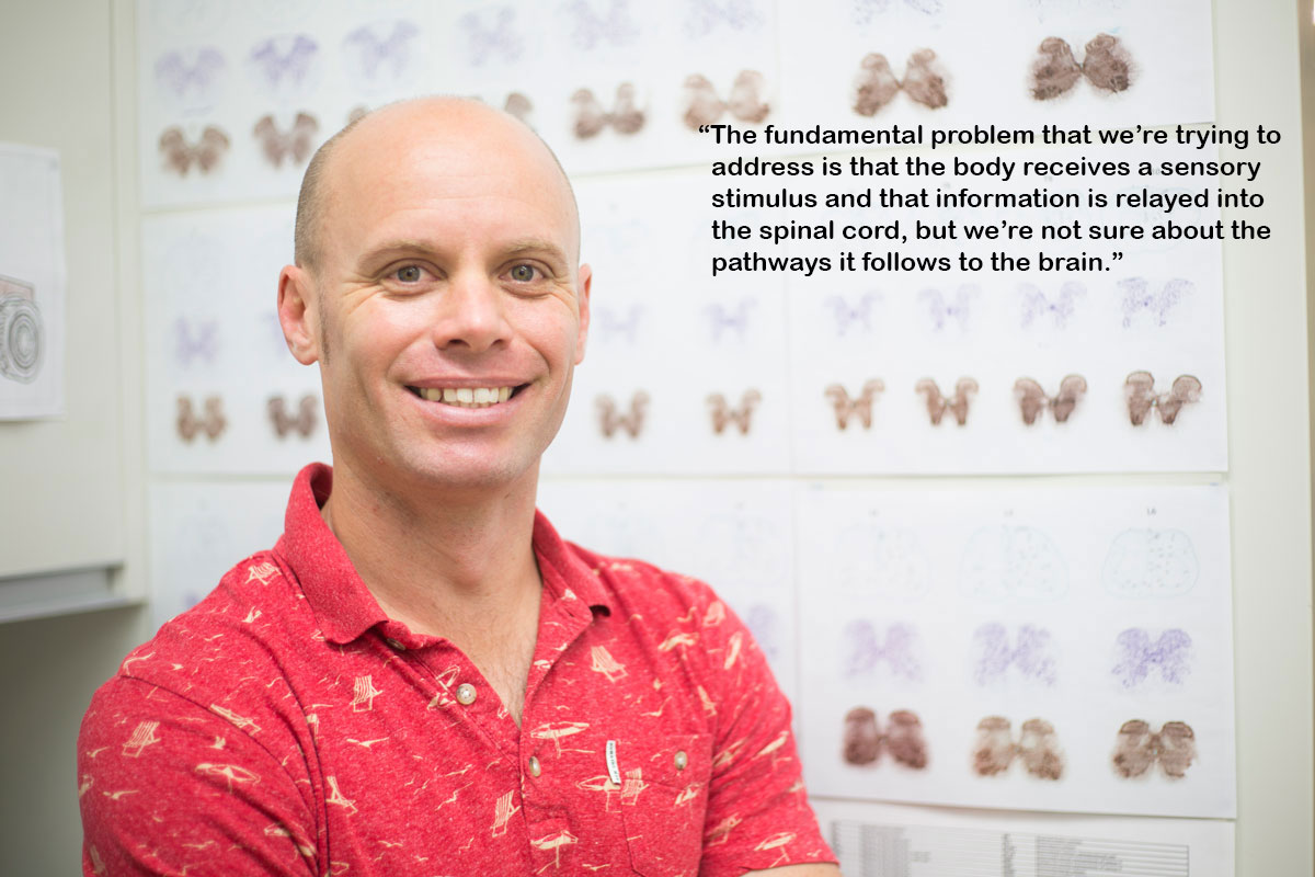
Projection neurons (PNs) in the Spinal Trigeminal Nucleus Caudalis (Sp5C) relay orofacial nociceptive information to higher brain regions such as the thalamus and the p... [more]
Projection neurons (PNs) in the Spinal Trigeminal Nucleus Caudalis (Sp5C) relay orofacial nociceptive information to higher brain regions such as the thalamus and the parabrachial nucleus (PBN). Our understanding of Sp5C PN organisation and function has advanced less than the parallel spinal cord output system despite their corresponding roles for transmission of nociceptive signals from the orofacial region and body respectively. Viral vectors are an established approach for studying circuit connectivity in the nervous system, but different serotypes are known to produce variable results across circuits. As such, we sought to validate the utility of two common viral serotypes in spinal PN research: retrograde adeno-associated virus serotype 2 (rgAAV) and adeno-associated virus serotype 9 (AAV9), for identifying and analysing Sp5C PNs that project to the PBN. Following unilateral injections of either viral serotype into the PBN, many Sp5C projection neurons were retrogradely labelled. For both serotypes, these injections labelled Sp5C PNs bilaterally with a strong bias to the ipsilateral Sp5C. Within Sp5C, similar levels of PN labelling were present in both superficial and deep regions, contrasting previous work in spinal PNs that showed greater labelling by AAV9 versus rgAAV. Comparisons of the age dependence of labelling showed greater retrograde labelling of Sp5C projection neurons when injections were made in young adult animals. Finally, we demonstrate successful Cre-dependent recombination to selectively express channelrhodopsin-2 in Sp5C projection neurons. Together, these experiments show that rgAAV and AAV9 produce strong Sp5C PN transduction and provide a basis for future study of the afferent and efferent functions of the Sp5C PN population in health and disease. (Figure presented.).
|
|
|
| 2025 |
Russo MA, Volschenk W, Bailey D, Santarelli DM, Holliday E, Barker D, Dizon J, Graham B, 'Twelve-Month Clinical Trial Results of a Novel, Dorsal Horn Dendrite Stimulation Waveform for Chronic Neuropathic Low Back Pain', Neuromodulation, 28, 263-273 (2025) [C1]
|
|
Open Research Newcastle |
| 2024 |
Russo MA, Santarelli DM, Austin PJ, Graham BA, 'Progressing into a new paradigm: how we must leave the past behind if we want a change in pain research outcomes', PAIN MEDICINE, 25, 5-7 (2024)
|
|
|
| 2024 |
Mitchell C, Campbell E, Fisher S, Stanton L, Burton N, Pearl A, Mcnally G, Bains J, Fuezesi T, Graham B, Manning E, Dayas C, 'Optogenetic recruitment of hypothalamic corticotrophin-releasing-hormone (CRH) neurons reduces motivational drive', TRANSLATIONAL PSYCHIATRY, 14 (2024) [C1]
|
|
Open Research Newcastle |
| 2024 |
Browne TJ, Smith KM, Gradwell MA, Dayas CV, Callister RJ, Hughes DI, Graham BA, 'Lateral lamina V projection neuron axon collaterals connect sensory processing across the dorsal horn of the mouse spinal cord', SCIENTIFIC REPORTS, 14 (2024) [C1]
|
|
Open Research Newcastle |
| 2024 |
Russo M, Santarelli D, Nash R, Graham B, 'ID: 329657 Rationale for Dorsal Horn Dendrite Stimulation', Neuromodulation Technology at the Neural Interface, 27 (2024)
|
|
|
| 2023 |
Davis OC, Dickie AC, Mustapa MB, Boyle KA, Browne TJ, Gradwell MA, Smith KM, Polgár E, Bell AM, Kókai É, Watanabe M, Wildner H, Zeilhofer HU, Ginty DD, Callister RJ, Graham BA, Todd AJ, Hughes DI, 'Calretinin-expressing islet cells: a source of pre- and post-synaptic inhibition of non-peptidergic nociceptor input to the mouse spinal cord.', bioRxiv (2023)
|
|
|
| 2023 |
Russo M, Volschenk W, Bailey D, Santarelli D, Graham B, 'ID: 210002 A Novel Spinal Cord Stimulation Waveform Targeting the Dorsal Horn: 12-Month Clinical Trial Results', Neuromodulation Technology at the Neural Interface, 26 (2023)
|
|
|
| 2023 |
Russo MA, Volschenk W, Bailey D, Santarelli DM, Holliday E, Barker D, Dizon J, Graham B, 'A Novel, Paresthesia-Free Spinal Cord Stimulation Waveform for Chronic Neuropathic Low Back Pain: Six-Month Results of a Prospective, Single-Arm, Dose-Response Study', NEUROMODULATION, 26, 1412-1423 (2023) [C1]
|
|
Open Research Newcastle |
| 2023 |
Russo M, Graham B, Santarelli DM, 'Gabapentin-Friend or foe?', PAIN PRACTICE, 23, 63-69 (2023) [C1]
|
|
Open Research Newcastle |
| 2023 |
Davis OC, Dickie AC, Mustapa MB, Boyle KA, Browne TJ, Gradwell MA, Smith KM, Polgar E, Bell AM, Kokai E, Watanabe M, Wildner H, Zeilhofer HU, Ginty DD, Callister RJ, Graham BA, Todd AJ, Hughes DI, 'Calretinin-expressing islet cells are a source of pre- and post-synaptic inhibition of non-peptidergic nociceptor input to the mouse spinal cord', SCIENTIFIC REPORTS, 13 (2023) [C1]
|
|
Open Research Newcastle |
| 2022 |
Iredale JA, Stoddard JG, Drury HR, Browne TJ, Elton A, Madden JF, Callister RJ, Welsh JS, Graham BA, 'Recording Network Activity in Spinal Nociceptive Circuits Using Microelectrode Arrays', Journal of Visualized Experiments
|
|
|
| 2022 |
Gradwell MA, Boyle KA, Browne TJ, Bell AM, Leonardo J, Reyes FSP, Dickie AC, Smith KM, Callister RJ, Dayas C, Hughes D, Graham BA, 'Diversity of inhibitory and excitatory parvalbumin interneuron circuits in the dorsal horn', PAIN, 163, E432-E452 (2022) [C1]
|
|
Open Research Newcastle |
| 2022 |
Iredale JA, Stoddard JG, Drury HR, Browne TJ, Elton A, Madden JF, Callister RJ, Welsh JS, Graham BA, 'Recording Network Activity in Spinal Nociceptive Circuits using Microelectrode Arrays', Journal of Visualized Experiments (2022) [C1]
|
|
Open Research Newcastle |
| 2022 |
Gradwell MA, Smith KM, Dayas CV, Smith DW, Hughes DI, Callister RJ, Graham BA, 'Altered Intrinsic Properties and Inhibitory Connectivity in Aged Parvalbumin-Expressing Dorsal Horn Neurons', FRONTIERS IN NEURAL CIRCUITS, 16 (2022) [C1]
|
|
Open Research Newcastle |
| 2021 |
Browne TJ, Smith KM, Gradwell MA, Iredale JA, Dayas CV, Callister RJ, Hughes DI, Graham BA, 'Spinoparabrachial projection neurons form distinct classes in the mouse dorsal horn', PAIN, 162, 1977-1994 (2021) [C1]
|
|
Open Research Newcastle |
| 2020 |
Madden JF, Davis OC, Boyle KA, Iredale JA, Browne TJ, Callister RJ, Smith DW, Jobling P, Hughes DI, Graham BA, 'Functional and Molecular Analysis of Proprioceptive Sensory Neuron Excitability in Mice', Frontiers in Molecular Neuroscience, 13, 1-13 (2020) [C1]
|
|
Open Research Newcastle |
| 2020 |
Callister RJ, Brichta AM, Schaefer AT, Graham BA, Stuart DG, 'Pioneers in CNS inhibition: 2. Charles Sherrington and John Eccles on inhibition in spinal and supraspinal structures', BRAIN RESEARCH, 1734 (2020) [C1]
|
|
Open Research Newcastle |
| 2020 |
Graham BA, Hughes DI, 'Defining populations of dorsal horn interneurons', PAIN, 161, 2434-2436 (2020)
|
|
|
| 2020 |
Gradwell MA, Callister RJ, Graham BA, 'Reviewing the case for compromised spinal inhibition in neuropathic pain', JOURNAL OF NEURAL TRANSMISSION, 127, 481-503 (2020) [C1]
A striking and debilitating property of the nervous system is that damage to this tissue can cause chronic intractable pain, which persists long after resolution of the... [more]
A striking and debilitating property of the nervous system is that damage to this tissue can cause chronic intractable pain, which persists long after resolution of the initial insult. This neuropathic form of pain can arise from trauma to peripheral nerves, the spinal cord, or brain. It can also result from neuropathies associated with disease states such as diabetes, human immunodeficiency virus/AIDS, herpes, multiple sclerosis, cancer, and chemotherapy. Regardless of the origin, treatments for neuropathic pain remain inadequate. This continues to drive research into the underlying mechanisms. While the literature shows that dysfunction in numerous loci throughout the CNS can contribute to chronic pain, the spinal cord and in particular inhibitory signalling in this region have remained major research areas. This review focuses on local spinal inhibition provided by dorsal horn interneurons, and how such inhibition is disrupted during the development and maintenance of neuropathic pain.
|
|
Open Research Newcastle |
| 2020 |
Browne TJ, Hughes DI, Dayas CV, Callister RJ, Graham BA, 'Projection Neuron Axon Collaterals in the Dorsal Horn: Placing a New Player in Spinal Cord Pain Processing', FRONTIERS IN PHYSIOLOGY, 11 (2020) [C1]
|
|
Open Research Newcastle |
| 2020 |
Mayhew JA, Cummins MJ, Cresswell ET, Callister RJ, Smith DW, Graham BA, 'Age-related gene expression changes in lumbar spinal cord: Implications for neuropathic pain', MOLECULAR PAIN, 16 (2020) [C1]
Clinically, pain has an uneven incidence throughout lifespan and impacts more on the elderly. In contrast, preclinical models of pathological pain have typically used j... [more]
Clinically, pain has an uneven incidence throughout lifespan and impacts more on the elderly. In contrast, preclinical models of pathological pain have typically used juvenile or young adult animals to highlight the involvement of glial populations, proinflammatory cytokines, and chemokines in the onset and maintenance of pathological signalling in the spinal dorsal horn. The potential impact of this mismatch is also complicated by the growing appreciation that the aged central nervous system exists in a state of chronic inflammation because of enhanced proinflammatory cytokine/chemokine signalling and glial activation. To address this issue, we investigated the impact of aging on the expression of genes that have been associated with neuropathic pain, glial signalling, neurotransmission and neuroinflammation. We used qRT-PCR to quantify gene expression and focussed on the dorsal horn of the spinal cord as this is an important perturbation site in neuropathic pain. To control for global vs region-specific age-related changes in gene expression, the ventral half of the spinal cord was examined. Our results show that expression of proinflammatory chemokines, pattern recognition receptors, and neurotransmitter system components was significantly altered in aged (24¿32 months) versus young mice (2¿4 months). Notably, the magnitude and direction of these changes were spinal-cord region dependent. For example, expression of the chemokine, Cxcl13, increased 119-fold in dorsal spinal cord, but only 2-fold in the ventral spinal cord of old versus young mice. Therefore, we propose the dorsal spinal cord of old animals is subject to region-specific alterations that prime circuits for the development of pathological pain, potentially in the absence of the peripheral triggers normally associated with these conditions.
|
|
Open Research Newcastle |
| 2020 |
Browne TJ, Gradwell MA, Iredale JA, Maden JF, Callister RJ, Hughes DI, Dayas CV, Graham BA, 'Transgenic Cross-Referencing of Inhibitory and Excitatory Interneuron Populations to Dissect Neuronal Heterogeneity in the Dorsal Horn', Frontiers in Molecular Neuroscience, 13, 1-20 (2020) [C1]
|
|
Open Research Newcastle |
| 2019 |
Yeoh JW, James MH, Adams CD, Bains JS, Sakurai T, Aston-Jones G, Graham BA, Dayas CV, 'Activation of lateral hypothalamic group III metabotropic glutamate receptors suppresses cocaine-seeking following abstinence and normalizes drug-associated increases in excitatory drive to orexin/hypocretin cells', NEUROPHARMACOLOGY, 154, 22-33 (2019) [C1]
The perifornical/lateral hypothalamic area (LHA) orexin (hypocretin) system is involved in drug-seeking behavior elicited by drug-associated stimuli. Cocaine exposure i... [more]
The perifornical/lateral hypothalamic area (LHA) orexin (hypocretin) system is involved in drug-seeking behavior elicited by drug-associated stimuli. Cocaine exposure is associated with presynaptic plasticity at LHA orexin cells such that excitatory input to orexin cells is enhanced acutely and into withdrawal. These changes may augment orexin cell reactivity to drug-related cues during abstinence and contribute to relapse-like behavior. Studies in hypothalamic slices from drug-naïve animals indicate that agonism of group III metabotropic glutamate receptors (mGluRs) reduces presynaptic glutamate release onto orexin cells. Therefore, we examined the group III mGluR system as a potential target to reduce orexin cell excitability in-vivo, including in animals with cocaine experience. First, we verified that group III mGluRs regulate orexin cell activity in behaving animals by showing that intra-LHA infusions of the selective agonist L-(+)-2-Amino-4-phosphonobutyric acid (L-AP4) reduces c-fos expression in orexin cells following 24 h food deprivation. Next, we extended these findings to show that intra-LHA L-AP4 infusions reduced discriminative stimulus-driven cocaine-seeking following withdrawal. Importantly, L-AP4 had no effect on lever pressing for sucrose pellets or general motoric behavior. Finally, using whole-cell patch-clamp recordings from identified orexin cells in orexin-GFP transgenic mice, we show enhanced presynaptic drive to orexin cells following 14d withdrawal and that this plasticity can be normalized by L-AP4. Together, these data indicate that activation of group III mGluRs in LHA reduces orexin cell activity in vivo and may be an effective strategy to suppress cocaine-seeking behavior following withdrawal. These effects are likely mediated, at least in part, by normalization of presynaptic plasticity at orexin cells that occurs as a result of cocaine exposure. This article is part of the Special Issue entitled 'Hypothalamic Control of Homeostasis'.
|
|
Open Research Newcastle |
| 2019 |
Mayhew JA, Callister RJ, Walker FR, Smith DW, Graham BA, 'Aging alters signaling properties in the mouse spinal dorsal horn', MOLECULAR PAIN, 15 (2019) [C1]
|
|
Open Research Newcastle |
| 2019 |
Boyle KA, Gradwell MA, Yasaka T, Dickie LC, Polgar E, Ganley RP, Orr DPH, Watanabe M, Abraira VE, Kuehn ED, Zimmerman AL, Ginty DD, Callister RJ, Graham BA, Hughes D, 'Defining a Spinal Microcircuit that Gates Myelinated Afferent Input: Implications for Tactile Allodynia', CELL REPORTS, 28, 526-+ (2019) [C1]
Chronic pain presents a major unmet clinical problem. The development of more effective treatments is hindered by our limited understanding of the neuronal circuits und... [more]
Chronic pain presents a major unmet clinical problem. The development of more effective treatments is hindered by our limited understanding of the neuronal circuits underlying sensory perception. Here, we show that parvalbumin (PV)-expressing dorsal horn interneurons modulate the passage of sensory information conveyed by low-threshold mechanoreceptors (LTMRs) directly via presynaptic inhibition and also gate the polysynaptic relay of LTMR input to pain circuits by inhibiting lamina II excitatory interneurons whose axons project into lamina I. We show changes in the functional properties of these PV interneurons following peripheral nerve injury and that silencing these cells unmasks a circuit that allows innocuous touch inputs to activate pain circuits by increasing network activity in laminae I¿IV. Such changes are likely to result in the development of tactile allodynia and could be targeted for more effective treatment of mechanical pain. In this study, Boyle et al. identify parvalbumin-expressing spinal interneurons as a principal source of axoaxonic synapses onto cutaneous myelinated afferents and inhibitory inputs onto lamina II vertical cells. Following peripheral nerve injury, disinhibition of these targets facilitates the aberrant recruitment of pain circuits, leading to tactile allodynia.
|
|
Open Research Newcastle |
| 2019 |
Ip CK, Zhang L, Farzi A, Qi Y, Clarke I, Reed F, Shi Y-C, Enriquez R, Dayas C, Graham B, Begg D, Bruening JC, Lee NJ, Hernandez-Sanchez D, Gopalasingam G, Koller J, Tasan R, Sperk G, Herzog H, 'Amygdala NPY Circuits Promote the Development of Accelerated Obesity under Chronic Stress Conditions', CELL METABOLISM, 30, 111-+ (2019) [C1]
Neuropeptide Y (NPY) exerts a powerful orexigenic effect in the hypothalamus. However, extra-hypothalamic nuclei also produce NPY, but its influence on energy homeostas... [more]
Neuropeptide Y (NPY) exerts a powerful orexigenic effect in the hypothalamus. However, extra-hypothalamic nuclei also produce NPY, but its influence on energy homeostasis is unclear. Here we uncover a previously unknown feeding stimulatory pathway that is activated under conditions of stress in combination with calorie-dense food; NPY neurons in the central amygdala are responsible for an exacerbated response to a combined stress and high-fat-diet intervention. Central amygdala NPY neuron-specific Npy overexpression mimics the obese phenotype seen in a combined stress and high-fat-diet model, which is prevented by the selective ablation of Npy. Using food intake and energy expenditure as readouts, we demonstrate that selective activation of central amygdala NPY neurons results in increased food intake and decreased energy expenditure. Mechanistically, it is the diminished insulin signaling capacity on central amygdala NPY neurons under combined stress and high-fat-diet conditions that leads to the exaggerated development of obesity.
|
|
Open Research Newcastle |
| 2019 |
Graham BA, Hughes DI, 'Rewards, perils and pitfalls of untangling spinal pain circuits', CURRENT OPINION IN PHYSIOLOGY, 11, 35-41 (2019) [C1]
Pain is a complex perception that is fundamental to our daily survival. Under normal circumstances, it serves an important protective function to guard against tissue d... [more]
Pain is a complex perception that is fundamental to our daily survival. Under normal circumstances, it serves an important protective function to guard against tissue damage or alert the body to dangerous environments. Under pathological states, however, the perception of pain can become chronic, maladaptive, resistant to treatment, and presents a serious clinical and societal problem. A wealth of literature suggests that disruption of sensory processing within the spinal cord contributes to chronic pain, but our limited understanding of spinal circuitry in health and disease remains a barrier to the development of new therapeutic strategies. The aim of this brief review is to outline current thinking about how individual components of functionally distinct spinal microcircuits can be identified and manipulated to determine their role in influencing our perception of pain in acute and chronic states.
|
|
Open Research Newcastle |
| 2019 |
Smith KM, Browne TJ, Davis OC, Coyle A, Boyle KA, Watanabe M, Dickinson SA, Iredale JA, Gradwell MA, Jobling P, Callister RJ, Dayas CV, Hughes DI, Graham BA, 'Calretinin positive neurons form an excitatory amplifier network in the spinal cord dorsal horn', ELIFE, 8 (2019) [C1]
|
|
Open Research Newcastle |
| 2018 |
Mayhew J, Graham BA, Biber K, Nilsson M, Walker FR, 'Purinergic modulation of glutamate transmission: An expanding role in stress-linked neuropathology', NEUROSCIENCE AND BIOBEHAVIORAL REVIEWS, 93, 26-37 (2018) [C1]
|
|
Open Research Newcastle |
| 2018 |
Tadros MA, Graham BA, Callister RJ, 'Moving functional classification of dorsal horn neurons from art to science', JOURNAL OF PHYSIOLOGY-LONDON, 596, 1543-1544 (2018)
|
|
|
| 2017 |
Gradwell MA, Boyle KA, Callister RJ, Hughes DI, Graham BA, 'Heteromeric /glycine receptors regulate excitability in parvalbumin-expressing dorsal horn neurons through phasic and tonic glycinergic inhibition', JOURNAL OF PHYSIOLOGY-LONDON, 595, 7185-7202 (2017) [C1]
Key points: Spinal parvalbumin-expressing interneurons have been identified as a critical source of inhibition to regulate sensory thresholds by gating mechanical input... [more]
Key points: Spinal parvalbumin-expressing interneurons have been identified as a critical source of inhibition to regulate sensory thresholds by gating mechanical inputs in the dorsal horn. This study assessed the inhibitory regulation of the parvalbumin-expressing interneurons, showing that synaptic and tonic glycinergic currents dominate, blocking neuronal or glial glycine transporters enhances tonic glycinergic currents, and these manipulations reduce excitability. Synaptically released glycine also enhanced tonic glycinergic currents and resulted in decreased parvalbumin-expressing interneuron excitability. Analysis of the glycine receptor properties mediating inhibition of parvalbumin neurons, as well as single channel recordings, indicates that heteromeric a/ß subunit-containing receptors underlie both synaptic and tonic glycinergic currents. Our findings indicate that glycinergic inhibition provides critical control of excitability in parvalbumin-expressing interneurons in the dorsal horn and represents a pharmacological target to manipulate spinal sensory processing. Abstract: The dorsal horn (DH) of the spinal cord is an important site for modality-specific processing of sensory information and is essential for contextually relevant sensory experience. Parvalbumin-expressing inhibitory interneurons (PV+ INs) have functional properties and connectivity that enables them to segregate tactile and nociceptive information. Here we examine inhibitory drive to PV+ INs using targeted patch-clamp recording in spinal cord slices from adult transgenic mice that express enhanced green fluorescent protein in PV+ INs. Analysis of inhibitory synaptic currents showed glycinergic transmission is the dominant form of phasic inhibition to PV+ INs. In addition, PV+ INs expressed robust glycine-mediated tonic currents; however, we found no evidence for tonic GABAergic currents. Manipulation of extracellular glycine by blocking either, or both, the glial and neuronal glycine transporters markedly decreased PV+ IN excitability, as assessed by action potential discharge. This decreased excitability was replicated when tonic glycinergic currents were increased by electrically activating glycinergic synapses. Finally, we show that both phasic and tonic forms of glycinergic inhibition are mediated by heteromeric a/ß glycine receptors. This differs from GABAA receptors in the dorsal horn, where different receptor stoichiometries underlie phasic and tonic inhibition. Together these data suggest both phasic and tonic glycinergic inhibition regulate the output of PV+ INs and contribute to the processing and segregation of tactile and nociceptive information. The shared stoichiometry for phasic and tonic glycine receptors suggests pharmacology is unlikely to be able to selectively target each form of inhibition in PV+ INs.
|
|
Open Research Newcastle |
| 2017 |
Farrell KE, Keely S, Walker MM, Brichta AM, Graham BA, Callister RJ, 'ALTERED INTRINSIC AND SYNAPTIC PROPERTIES OF LUMBOSACRAL DORSAL HORN NEURONS IN A MOUSE MODEL OF COLITIS', NEUROSCIENCE, 362, 152-167 (2017) [C1]
Visceral pain in inflammatory and functional gastrointestinal conditions is a major clinical problem. The exact mechanisms underlying the development of pain, during an... [more]
Visceral pain in inflammatory and functional gastrointestinal conditions is a major clinical problem. The exact mechanisms underlying the development of pain, during and after visceral inflammation are unknown. However, clinical and pre-clinical evidence suggests plasticity within the spinal cord dorsal horn is a contributing factor. Here we use an in vivo preparation and patch-clamp electrophysiology to test whether the synaptic and intrinsic properties of superficial dorsal horn (SDH) neurons are altered 5 days after the induction of mild colitis in adult male mice (i.e. during acute inflammation of the colon). Whole-cell recordings were made from lumbosacral (L6-S1) superficial dorsal horn neurons (SDH), in animals under isoflurane anesthesia. Noxious colorectal distension (CRD) was used to identify SDH neurons with colonic inputs, while stimulation of the hind paw and tail was employed to assess convergent cutaneous input. Following inflammation, a significantly increased proportion of SDH neurons received both colonic and cutaneous inputs, compared to neurons in naïve animals. In addition, the nature and magnitude of responses to CRD and cutaneous stimulation differed in inflamed animals, as was spontaneous excitatory synaptic drive. Conversely, several measures of intrinsic excitability were altered in a manner that would decrease SDH network excitability following colitis. We propose that during inflammation, sensitization of colonic afferents results in increased signaling to the SDH. This is accompanied by plasticity in SDH neurons whereby their intrinsic properties are changed to compensate for altered afferent activity.
|
|
Open Research Newcastle |
| 2017 |
Campbell EJ, Mitchell CS, Adams CD, Yeoh JW, Hodgson DM, Graham BA, Dayas CV, 'Chemogenetic activation of the lateral hypothalamus reverses early life stress-induced deficits in motivational drive', EUROPEAN JOURNAL OF NEUROSCIENCE, 46, 2285-2296 (2017) [C1]
Altered motivated behaviour is a cardinal feature of several neuropsychiatric conditions including mood disorders. One well-characterized antecedent to the development ... [more]
Altered motivated behaviour is a cardinal feature of several neuropsychiatric conditions including mood disorders. One well-characterized antecedent to the development of mood disorders is exposure to early life stress (ELS). A key brain substrate controlling motivated behaviour is the lateral hypothalamus (LH). Here, we examined the effect of ELS on LH activation and the motivation to self-administer sucrose. We tested whether chemogenetic activation of LH circuits could modify sucrose responding in ELS rats and examined the impact on LH cell populations. Male rat pups were maternally separated for 0 or 3¿h on postnatal days 2¿14. During adolescence, rats received bilateral injections of hM3D(Gq), the excitatory designer receptor exclusively activated by designer drugs, into LH. In adulthood, rats were trained to self-administer sucrose and tested under a progressive ratio schedule to determine their motivation for reward following injection with either vehicle or 5¿mg/kg clozapine-N-oxide. Brains were processed for Fos-protein immunohistochemistry. ELS significantly suppressed lever responding for sucrose, indicating a long-lasting impact of ELS on motivation circuits. hM3D(Gq) activation of LH increased responding, normalizing deficits in ELS rats, and increased Fos-positive orexin and MCH cell numbers within LH. Our findings indicate that despite being susceptible to environmental stressors, LH circuits retain the capacity to overcome ELS-induced deficits in motivated behaviour.
|
|
Open Research Newcastle |
| 2017 |
Tae H-S, Smith KM, Phillips AM, Boyle KA, Li M, Forster IC, Hatch RJ, Richardson R, Hughes DI, Graham BA, Petrou S, Reid CA, 'Gabapentin Modulates HCN4 Channel Voltage-Dependence', FRONTIERS IN PHARMACOLOGY, 8 (2017) [C1]
|
|
Open Research Newcastle |
| 2017 |
Flynn JR, Conn VL, Boyle KA, Hughes DI, Watanabe M, Velasquez T, Goulding MD, Callister RJ, Graham BA, 'Anatomical and Molecular Properties of Long Descending Propriospinal Neurons in Mice', FRONTIERS IN NEUROANATOMY, 11 (2017) [C1]
|
|
Open Research Newcastle |
| 2017 |
Huisman MV, Rothman KJ, Paquette M, Teutsch C, Diener H-C, Dubner SJ, Halperin JL, Ma CS, Zint K, Elsaesser A, Bartels DB, Lip GYH, Abban D, Abdul N, Abelson M, Ackermann A, Adams F, Adams L, Adragao P, Ageno W, Aggarwal R, Agosti S, Marin JA, Aguilar F, Linares JAA, Aguinaga L, Ahmad Z, Ainsworth P, Al Ghalayini K, Al Ismail S, Alasfar A, Alawwa A, Al-Dallow R, Alderson L, Alexopoulos D, Ali A, Ali M, Aliyar P, Al-Joundi T, Al Mahameed S, Almassi H, Almuti K, Al-Obaidi M, Alshehri M, Altmann U, Alves AR, Al-Zoebi A, Amara W, Amelot M, Amjadi N, Ammirati F, Andrawis N, Angoulvant D, Annoni G, Ansalone G, Antonescu SA, Ariani M, Arias JC, Armero S, Arora R, Arora C, Ashcraft W, Aslam MS, Astesiano A, Audouin P, Augenbraun C, Aydin S, Azar R, Azim A, Aziz S, Backes LM, Baig M, Bains S, Bakbak A, Baker S, Bakhtiar K, Bala R, Banayan J, Bandh S, Bando S, Banerjee S, Bank A, Barbarash O, Baron G, Barr C, Barrera C, Barton J, Kes VB, Baula G, Bayeh H, Bazargani N, Behrens S, Bell A, Benezet-Mazuecos J, Benhalima B, Berdague P, van den Berg BJ, van Bergen PFMM, Berngard E, Bernstein R, Berrospi P, Berti S, Bertomeu V, Berz A, Bettencourt P, Betzu R, Beyer-Westendorf J, Bhagwat R, Black T, Ibaceta JHB, Bloom S, Blumberg E, Bo M, Bockisch V, Bohmer E, Bongiorni MG, Boriani G, Bosch R, Boswijk DJ, Bott J, Bottacchi E, Kalan MB, Brandes A, Bratland B, Brautigam D, Breton N, Brouwers PJAM, Browne K, Bruguera J, Brunehaut M, Brunschwig C, Buathier H, Buhl A, Bullinga J, Butcher K, Honorio JWC, Caccavo A, Cadinot D, Cai S, Calvi V, Camm J, Candeias R, Capo J, Capucci A, Cardoso JN, Vera YCD, Carlson B, Carvalho P, Cary S, Casanova R, Casu G, Cattan S, Cavallini C, Cayla G, Cha TJ, Cha KS, Chaaban S, Chae JK, Challappa K, Chand S, Chandrashekar H, Chang M, Charbel P, Chartier L, Chatterjee K, Cheema A, Chen S-A, Chevallereau P, Chiang F-T, Chiarella F, Lin C-C, Cho YK, Choi DJ, Chouinard G, Danny HFC, Chrysos D, Chumakova G, Roberto EJJ, Valenzuela C, Cieza-Lara T, Nica VC, Ciobot
|
|
|
| 2016 |
Duchatel RJ, Jobling P, Graham BA, Harms LR, Michie PT, Hodgson DM, Tooney PA, 'Increased white matter neuron density in a rat model of maternal immune activation - Implications for schizophrenia', PROGRESS IN NEURO-PSYCHOPHARMACOLOGY & BIOLOGICAL PSYCHIATRY, 65 (2016) [C1]
Interstitial neurons are located among white matter tracts of the human and rodent brain. Post-mortem studies have identified increased interstitial white matter neuron... [more]
Interstitial neurons are located among white matter tracts of the human and rodent brain. Post-mortem studies have identified increased interstitial white matter neuron (IWMN) density in the fibre tracts below the cortex in people with schizophrenia. The current study assesses IWMN pathology in a model of maternal immune activation (MIA); a risk factor for schizophrenia. Experimental MIA was produced by an injection of polyinosinic:polycytidylic acid (PolyI:C) into pregnant rats on gestational day (GD) 10 or GD19. A separate control group received saline injections. The density of neuronal nuclear antigen (NeuN<sup>+</sup>) and somatostatin (SST<sup>+</sup>) IWMNs was determined in the white matter of the corpus callosum in two rostrocaudally adjacent areas in the 12week old offspring of GD10 (n=10) or GD19 polyI:C dams (n=18) compared to controls (n=20). NeuN<sup>+</sup> IWMN density trended to be higher in offspring from dams exposed to polyI:C at GD19, but not GD10. A subpopulation of these NeuN<sup>+</sup> IWMNs was shown to express SST. PolyI:C treatment of dams induced a significant increase in the density of SST<sup>+</sup> IWMNs in the offspring when delivered at both gestational stages with more regionally widespread effects observed at GD19. A positive correlation was observed between NeuN<sup>+</sup> and SST<sup>+</sup> IWMN density in animals exposed to polyI:C at GD19, but not controls. This is the first study to show that MIA increases IWMN density in adult offspring in a similar manner to that seen in the brain in schizophrenia. This suggests the MIA model will be useful in future studies aimed at probing the relationship between IWMNs and schizophrenia.
|
|
Open Research Newcastle |
| 2016 |
Farrell KE, Rank MM, Keely S, Brichta AM, Graham BA, Callister RJ, 'IN VIVO CHARACTERIZATION OF COLORECTAL AND CUTANEOUS INPUTS TO LUMBOSACRAL DORSAL HORN NEURONS IN THE MOUSE SPINAL CORD', NEUROSCIENCE, 316, 13-25 (2016) [C1]
Chronic abdominal pain is a common symptom of inflammatory bowel disease and often persists in the absence of gut inflammation. Although the mechanisms responsible for ... [more]
Chronic abdominal pain is a common symptom of inflammatory bowel disease and often persists in the absence of gut inflammation. Although the mechanisms responsible for ongoing pain are unknown, clinical and preclinical evidence suggests lumbosacral spinal cord dorsal horn neurons contribute to these symptoms. At present, we know little about the intrinsic and synaptic properties of this population of neurons in either normal or inflammed conditions. Therefore, we developed an in vivo preparation to make patch-clamp recordings from superficial dorsal horn (SDH) neurons receiving colonic inputs in naïve male mice. Recordings were made in the lumbosacral spinal cord (L6-S1) under isoflurane anesthesia. Noxious colorectal distension (CRD) was used to determine whether SDH neurons received inputs from mechanical stimulation/distension of the colon. Responses to hind paw/tail cutaneous stimulation and intrinsic and synaptic properties were also assessed, as well as action potential discharge properties. Approximately 11% of lumbosacral SDH neurons in the cohort of neurons sampled responded to CRD and a majority of these responses were subthreshold. Most CRD-responsive neurons (80%) also responded to cutaneous stimuli, compared with <50% of CRD-non-responsive neurons. Furthermore, CRD-responsive neurons had more hyperpolarized resting membrane potentials, larger rheobase currents, and reduced levels of excitatory drive, compared to CRD-non-responsive neurons. Our results demonstrate that CRD-responsive neurons can be distinguished from CRD-non-responsive neurons by several differences in their membrane properties and excitatory synaptic inputs. We also demonstrate that SDH neurons with colonic inputs show predominately subthreshold responses to CRD and exhibit a high degree of viscerosomatic convergence.
|
|
Open Research Newcastle |
| 2016 |
Smith KM, Boyle KA, Mustapa M, Jobling P, Callister RJ, Hughes DI, Graham BA, 'DISTINCT FORMS OF SYNAPTIC INHIBITION AND NEUROMODULATION REGULATE CALRETININ-POSITIVE NEURON EXCITABILITY IN THE SPINAL CORD DORSAL HORN', NEUROSCIENCE, 326, 19-30 (2016) [C1]
The dorsal horn (DH) of the spinal cord contains a heterogenous population of neurons that process incoming sensory signals before information ascends to the brain. We ... [more]
The dorsal horn (DH) of the spinal cord contains a heterogenous population of neurons that process incoming sensory signals before information ascends to the brain. We have recently characterized calretinin-expressing (CR+) neurons in the DH and shown that they can be divided into excitatory and inhibitory subpopulations. The excitatory population receives high-frequency excitatory synaptic input and expresses delayed firing action potential discharge, whereas the inhibitory population receives weak excitatory drive and exhibits tonic or initial bursting discharge. Here, we characterize inhibitory synaptic input and neuromodulation in the two CR+ populations, in order to determine how each is regulated. We show that excitatory CR+ neurons receive mixed inhibition from GABAergic and glycinergic sources, whereas inhibitory CR+ neurons receive inhibition, which is dominated by glycine. Noradrenaline and serotonin produced robust outward currents in excitatory CR+ neurons, predicting an inhibitory action on these neurons, but neither neuromodulator produced a response in CR+ inhibitory neurons. In contrast, enkephalin (along with selective mu and delta opioid receptor agonists) produced outward currents in inhibitory CR+ neurons, consistent with an inhibitory action but did not affect the excitatory CR+ population. Our findings show that the pharmacology of inhibitory inputs and neuromodulator actions on CR+ cells, along with their excitatory inputs can define these two subpopulations further, and this could be exploited to modulate discrete aspects of sensory processing selectively in the DH.
|
|
Open Research Newcastle |
| 2016 |
Whelan KR, Hamilton J, Peeling L, Graham B, Hunter G, Kelly ME, 'Importance of Developing Stroke Systems of Care to Improve Access to Endovascular Therapies', WORLD NEUROSURGERY, 88, 678-680 (2016)
|
|
|
| 2015 |
Kongsui R, Johnson SJ, Graham BA, Nilsson M, Walker FR, 'A COMBINED CUMULATIVE THRESHOLD SPECTRA AND DIGITAL RECONSTRUCTION ANALYSIS REVEAL STRUCTURAL ALTERATIONS OF MICROGLIA WITHIN THE PREFRONTAL CORTEX FOLLOWING LOW-DOSE LPS ADMINISTRATION', NEUROSCIENCE, 310, 629-640 (2015) [C1]
Sickness behaviors have become the focus of great interest in recent years as they represent a clear case of how peripheral disturbances in immune signaling can disrupt... [more]
Sickness behaviors have become the focus of great interest in recent years as they represent a clear case of how peripheral disturbances in immune signaling can disrupt quite complex behaviors. In the current study, we were interested in examining whether we could identify any significant morphological disturbances in microglia associated with these sickness-like behaviors in adult male Sprague-Dawley rats. We chose lipopolysaccharide (LPS 100 µg/kg/i.p.), to induce sickness-like behaviors as it is the most well-validated approach to do so in rodents and humans. We were particularly interested in examining changes in microglia within the prefrontal cortex (PFC) as several recent neuroimaging studies have highlighted significant functional changes in this region following peripheral LPS administration. Paraformaldehyde-fixed tissue was collected from animals 24 h post LPS administration and labeled immunohistochemically with an antibody directed to bind to Iba-1, a protein known to be involved in the structural remodeling of microglia. To analyze changes, we have made use of two recently described image analysis procedures. The first is known as cumulative threshold spectra (CTS) analysis. The second involves the unsupervised digital reconstruction of microglia. We undertook these complementary analysis of microglial cells in the both the pre- and infralimbic divisions of the PFC. Our results indicated that microglial soma size was significantly enlarged, while cell processes had contracted slightly following LPS administration. To our knowledge this study is to first to definitely demonstrate substantial microglial disturbances within the PFC following LPS delivered at a dose that was sufficient to induce significant sickness-like behavior.
|
|
Open Research Newcastle |
| 2015 |
Tadros MA, Farrell KE, Graham BA, Brichta AM, Callister RJ, 'Properties of sodium currents in neonatal and young adult mouse superficial dorsal horn neurons.', Molecular pain, 11 (2015)
|
|
|
| 2015 |
Callister RJ, Graham BA, 'Spicing up the gabapentionoids: Facilitating gabapentin entry in spinal pain circuits', NEUROSCIENCE LETTERS, 584, 395-396 (2015) [C3]
|
|
|
| 2015 |
Smith KM, Boyle KA, Madden JF, Dickinson SA, Jobling P, Callister RJ, Hughes DI, Graham BA, 'Functional heterogeneity of calretinin-expressing neurons in the mouse superficial dorsal horn: implications for spinal pain processing', JOURNAL OF PHYSIOLOGY-LONDON, 593, 4319-4339 (2015) [C1]
Neurons in the superficial dorsal horn (SDH) of the spinal cord play an important role in nociceptive, thermal, itch and light touch sensations. Excitatory interneurons... [more]
Neurons in the superficial dorsal horn (SDH) of the spinal cord play an important role in nociceptive, thermal, itch and light touch sensations. Excitatory interneurons comprise ~65% of all SDH neurons but surprisingly few studies have investigated their role in spinal sensory processing. Here we use a transgenic mouse to study putative excitatory SDH neurons that express the calcium binding protein calretinin (CR). Our immunocytochemical, morphological and electrophysiological analysis identified two distinct populations of CR-expressing neurons, which we termed 'Typical' and 'Atypical'. Typical CR-expressing neurons comprised ~85% of the population and exhibited characteristic excitatory interneuron properties including delayed firing discharge, large rapid A-type potassium currents, and central, radial or vertical cell morphologies. Atypical neurons exhibited properties consistent with inhibitory interneurons, including tonic firing or initial bursting discharge, Ih currents, and islet cell morphology. Although both Typical and Atypical CR-expressing neurons responded to noxious peripheral stimulation, the excitatory drive onto Typical CR-expressing neurons was much stronger. Furthermore, Atypical CR-expressing cells comprise at least two functionally distinct subpopulations based on their responsiveness to noxious peripheral stimulation and neurochemical profile. Together our data suggest CR expression is not restricted to excitatory neurons in the SDH. Under normal conditions, the contribution of 'Typical' excitatory CR-expressing neurons to overall SDH excitability may be limited by the presence of A-type potassium currents, which limit the effectiveness of their strong excitatory input. Their contribution may, however, be increased in pathological situations where A-type potassium currents are decreased. By contrast, 'Atypical' inhibitory neurons with their excitable phenotype but weak excitatory input may be more easily recruited during increased peripheral stimulation.
|
|
Open Research Newcastle |
| 2014 |
Tadros MA, Farrell KE, Schofield PR, Brichta AM, Graham BA, Fuglevand AJ, Callister RJ, 'Intrinsic and synaptic homeostatic plasticity in motoneurons from mice with glycine receptor mutations', JOURNAL OF NEUROPHYSIOLOGY, 111, 1487-1498 (2014) [C1]
Inhibitory synaptic inputs to hypoglossal motoneurons (HMs) are important for modulating excitability in brainstem circuits. Here we ask whether reduced inhibition, as ... [more]
Inhibitory synaptic inputs to hypoglossal motoneurons (HMs) are important for modulating excitability in brainstem circuits. Here we ask whether reduced inhibition, as occurs in three murine mutants with distinct naturally occurring mutations in the glycine receptor (GlyR), leads to intrinsic and/or synaptic homeostatic plasticity. Whole cell recordings were obtained from HMs in transverse brainstem slices from wild-type (wt), spasmodic (spd), spastic (spa), and oscillator (ot) mice (C57Bl/6, approximately postnatal day 21). Passive and action potential (AP) properties in spd and ot HMs were similar to wt. In contrast, spa HMs had lower input resistances, more depolarized resting membrane potentials, higher rheobase currents, smaller AP amplitudes, and slower afterhyperpolarization current decay times. The excitability of HMs, assessed by "gain" in injected current/firing-frequency plots, was similar in all strains whereas the incidence of rebound spiking was increased in spd. The difference between recruitment and derecruitment current (i.e., ¿I) for AP discharge during ramp current injection was more negative in spa and ot. GABAA miniature inhibitory postsynaptic current (mIPSC) amplitude was increased in spa and ot but not spd, suggesting diminished glycinergic drive leads to compensatory adjustments in the other major fast inhibitory synaptic transmitter system in these mutants. Overall, our data suggest long-term reduction in glycinergic drive to HMs results in changes in intrinsic and synaptic properties that are consistent with homeostatic plasticity in spa and ot but not in spd. We propose such plasticity is an attempt to stabilize HM output, which succeeds in spa but fails in ot. © 2014 the American Physiological Society.
|
|
Open Research Newcastle |
| 2014 |
Farrell KE, Keely S, Graham BA, Callister R, Callister RJ, 'A Systematic Review of the Evidence for Central Nervous System Plasticity in Animal Models of Inflammatory-mediated Gastrointestinal Pain', INFLAMMATORY BOWEL DISEASES, 20, 176-195 (2014) [C1]
|
|
Open Research Newcastle |
| 2014 |
Yeoh JW, Campbell EJ, James MH, Graham BA, Dayas CV, 'Orexin antagonists for neuropsychiatric disease: progress and potential pitfalls.', Frontiers in neuroscience, 8, 36-36 (2014) [C1]
|
|
Open Research Newcastle |
| 2014 |
Yeoh JW, James MH, Graham BA, Dayas CV, 'Electrophysiological characteristics of paraventricular thalamic (PVT) neurons in response to cocaine and cocaine- and amphetamine-regulated transcript (CART)', FRONTIERS IN BEHAVIORAL NEUROSCIENCE, 8 (2014) [C1]
|
|
Open Research Newcastle |
| 2014 |
Harris BM, Hughes DI, Bolton PS, Tadros MA, Callister RJ, Graham BA, 'Contrasting alterations to synaptic and intrinsic properties in upper-cervical superficial dorsal horn neurons following acute neck muscle inflammation', MOLECULAR PAIN, 10 (2014) [C1]
|
|
Open Research Newcastle |
| 2014 |
Stuart DG, Schaefer AT, Massion J, Graham BA, Callister RJ, 'Pioneers in CNS inhibition: 1. Ivan M. Sechenov, the first to clearly demonstrate inhibition arising in the brain', BRAIN RESEARCH, 1548, 20-48 (2014) [C1]
This article reviews the contributions of Ivan Michailovich Sechenov [1829-1905] to the neurophysiological concept of central inhibition. He first studied this concept ... [more]
This article reviews the contributions of Ivan Michailovich Sechenov [1829-1905] to the neurophysiological concept of central inhibition. He first studied this concept in the frog and on himself. Later his trainees extended the study of central inhibition to other mammalian species. Outside his own country, Sechenov is better known for his prescient contributions to physiological psychology. In Russia, however, he is also revered as "the father of Russian physiology," because of his contributions to neurophysiology and other aspects of physiology including blood gases and respiration, the physiology and biomechanics of movement, and general physiology concepts that appeared in his textbooks and later works he helped translate from largely German sources. After graduation from Moscow University Medical School in 1856 he spent 31/2 years in Germany and Austria where he attended lectures and conducted research under the direction of several prominent physiologists and biochemists. In his subsequent academic career he held positions at universities in St. Petersburg (1860-1870; 1876-1888), Odessa (1871-1876) and Moscow (1890-1905). From 1860 onwards he was acclaimed as a physiologist in academic circles. He was also well known in Russian society for his public lectures on physiology and his views on physiological psychology. The latter resulted in him being branded "politically unreliable" by the tsarist bureaucracy from 1863 onwards. Sechenov's first (1862) study on central inhibition remains his most memorable. He delayed the withdrawal of a frog's foot from a weak acid solution by chemical or electrical stimulation of selected parts of the central nervous system. He also noted similar effects on his own hand during co-activation of other sensory inputs by tickling or teeth gnashing. © 2013 Elsevier B.V. All rights reserved.
|
|
Open Research Newcastle |
| 2014 |
Smith KM, Madden JF, Callister RJ, Hughes DI, Graham BA, 'The search for novel analgesics: re-examining spinal cord circuits with new tools.', Frontiers in pharmacology, 5 (2014) [C1]
|
|
Open Research Newcastle |
| 2013 |
Hughes DI, Boyle KA, Kinnon CM, Bilsland C, Quayle JA, Callister RJ, Graham BA, 'HCN4 SUBUNIT EXPRESSION IN FAST-SPIKING INTERNEURONS OF THE RAT SPINAL CORD AND HIPPOCAMPUS', NEUROSCIENCE, 237, 7-18 (2013) [C1]
Hyperpolarisation-activated (Ih) currents are considered important for dendritic integration, synaptic transmission, setting membrane potential and rhythmic action pote... [more]
Hyperpolarisation-activated (Ih) currents are considered important for dendritic integration, synaptic transmission, setting membrane potential and rhythmic action potential (AP) discharge in neurons of the central nervous system. Hyperpolarisation-activated cyclic nucleotide-gated (HCN) channels underlie these currents and are composed of homo- and hetero-tetramers of HCN channel subunits (HCN1-4), which confer distinct biophysical properties on the channel. Despite understanding the structure-function relationships of HCN channels with different subunit stoichiometry, our knowledge of their expression in defined neuronal populations remains limited. Recently, we have shown that HCN subunit expression is a feature of a specific population of dorsal horn interneurons that exhibit high-frequency AP discharge. Here we expand on this observation and use neuroanatomical markers to first identify well-characterised neuronal populations in the lumbar spinal cord and hippocampus and subsequently determine whether HCN4 expression correlates with high-frequency AP discharge in these populations. In the spinal cord, HCN4 is expressed in several putative inhibitory interneuron populations including parvalbumin (PV)-expressing islet cells (84.1%; SD: ±2.87), in addition to all putative Renshaw cells and Ia inhibitory interneurons. Similarly, virtually all PV-expressing cells in the hippocampal CA1 subfield (93.5%; ±3.40) and the dentate gyrus (90.9%; ±6.38) also express HCN4. This HCN4 expression profile in inhibitory interneurons mirrors both the prevalence of Ih sub-threshold currents and high-frequency AP discharge. Our findings indicate that HCN4 subunits are expressed in several populations of spinal and hippocampal interneurons, which are known to express both Ih sub-threshold currents and exhibit high-frequency AP discharge. As HCN channel function plays a critical role in pain perception, learning and memory, and sleep as well as the pathogenesis of several neurological diseases, these findings provide important insights into the identity and neurochemical status of cells that could underlie such conditions. © 2013 IBRO.
|
|
Open Research Newcastle |
| 2012 |
James MH, Yeoh JW, Graham B, Dayas C, 'Insights for Developing Pharmacological Treatments for Psychostimulant Relapse Targeting Hypothalamic Peptide Systems.', Journal of Addiction Research and Therapy, 01, 1-14 (2012)
|
|
Open Research Newcastle |
| 2012 |
Tadros MA, Harris B, Anderson WB, Brichta AM, Graham BA, Callister RJ, 'Are all spinal segments equal: Intrinsic membrane properties of superficial dorsal horn neurons in the developing and mature mouse spinal cord', Journal of Physiology, 590, 2409-2425 (2012) [C1]
|
|
Open Research Newcastle |
| 2012 |
Yeoh JW, James MH, Jobling P, Bains JS, Graham BA, Dayas CV, 'Cocaine potentiates excitatory drive in the perifornical/lateral hypothalamus', Journal of Physiology, 590, 3677-3689 (2012) [C1]
|
|
Open Research Newcastle |
| 2012 |
Hughes DI, Sikander S, Kinnon CM, Boyle KA, Watanabe M, Callister RJ, Graham BA, 'Morphological, neurochemical and electrophysiological features of parvalbumin-expressing cells: A likely source of axo-axonic inputs in the mouse spinal dorsal horn', Journal of Physiology, 590, 3927-3951 (2012) [C1]
|
|
Open Research Newcastle |
| 2011 |
Graham BA, Tadros MA, Schofield PR, Callister RJ, 'Probing glycine receptor stoichiometry in superficial dorsal horn neurones using the spasmodic mouse', Journal of Physiology, 589, 2459-2474 (2011) [C1]
|
|
Open Research Newcastle |
| 2011 |
Flynn JR, Graham BA, Galea MP, Callister RJ, 'The role of propriospinal interneurons in recovery from spinal cord injury', Neuropharmacology, 60, 809-822 (2011) [C1]
|
|
Open Research Newcastle |
| 2011 |
Flynn JR, Brichta AM, Galea MP, Callister RJ, Graham BA, 'A horizontal slice preparation for examining the functional connectivity of dorsal column fibres in mouse spinal cord', Journal of Neuroscience Methods, 200, 113-120 (2011) [C1]
|
|
Open Research Newcastle |
| 2010 |
Callister RJ, Graham BA, 'Early history of glycine receptor biology in mammalian spinal cord circuits', Frontiers in Molecular Neuroscience, 3, 1-13 (2010) [C1]
|
|
Open Research Newcastle |
| 2010 |
Jobling P, Graham BA, Brichta AM, Callister RJ, 'Cervix stimulation evokes predominantly subthreshold synaptic responses in mouse thoracolumbar and lumbosacral superficial dorsal horn neurons', Journal of Sexual Medicine, 7, 2068-2076 (2010) [C1]
|
|
Open Research Newcastle |
| 2010 |
De Oliveira R, Graham BA, Howlett MC, Gravina FS, Oliveira MW, Imtiaz MS, Callister RJ, Lim R, Brichta AM, Van Helden DF, 'Ketamine anesthesia helps preserve neuronal viability', Journal of Neuroscience Methods, 189, 230-232 (2010) [C1]
|
|
Open Research Newcastle |
| 2010 |
Graham BA, Clausen PD, Bolton PS, 'A descriptive study of the force and displacement profiles of the toggle-recoil spinal manipulative procedure (adjustment) as performed by chiropractors', Manual Therapy, 15, 74-79 (2010) [C1]
|
|
Open Research Newcastle |
| 2009 |
Anderson WB, Graham BA, Beveridge NJ, Tooney PA, Brichta AM, Callister RJ, 'Different forms of glycine- and GABA(A)-receptor mediated inhibitory synaptic transmission in mouse superficial and deep dorsal horn neurons', Molecular Pain, 5, 1-16 (2009) [C1]
|
|
Open Research Newcastle |
| 2009 |
Tadros MA, Graham BA, Brichta AM, Callister RJ, 'Evidence for a critical period in the development of excitability and potassium currents in mouse lumbar superficial dorsal horn neurons', Journal of Neurophysiology, 101, 1800-1812 (2009) [C1]
|
|
Open Research Newcastle |
| 2008 |
Graham BA, Brichta AM, Callister RJ, 'Recording temperature affects the excitability of mouse superficial dorsal horn neurons, in vitro', Journal of Neurophysiology, 99, 2048-2059 (2008) [C1]
|
|
Open Research Newcastle |
| 2007 |
Graham BA, Brichta AM, Callister RJ, 'Moving from an averaged to specific view of spinal cord pain processing circuits', Journal of Neurophysiology, 98, 1057-1063 (2007) [C1]
|
|
Open Research Newcastle |
| 2007 |
Graham BA, Brichta AM, Callister RJ, 'Pinch-current injection defines two discharge profiles in mouse superficial dorsal horn neurones, in vitro', Journal of Physiology, 578, 787-798 (2007) [C1]
|
|
Open Research Newcastle |
| 2007 |
Graham BA, Brichta AM, Schofield PR, Callister RJ, 'Altered potassium channel function in the superficial dorsal horn of the spastic mouse', Journal of Physiology, 584, 121-136 (2007) [C1]
|
|
Open Research Newcastle |
| 2006 |
Graham BA, Schofield PR, Sah P, Margrie TW, Callister RJ, 'Distinct physiological mechanisms underlie altered glycinergic synaptic transmission in the murine mutants, spastic, spasmodic, and oscillator', Journal of Neuroscience, 26, 4880-4890 (2006) [C1]
|
|
Open Research Newcastle |
| 2004 |
Graham BA, Brichta AM, Callister RJ, 'In vivo responses of mouse superficial dorsal horn neurones to both current injection and peripheral cutaneous stimulation', Journal of Physiology, 561.3, 749-763 (2004) [C1]
|
|
Open Research Newcastle |
| 2004 |
Graham BA, Brichta AM, Callister RJ, 'An in vivo mouse spinal cord preparation for patch-clamp analysis of nociceptive processing', Journal of Neuroscience Methods, 136, 221-228 (2004) [C1]
|
|
|
| 2003 |
Graham BA, Schofield PR, Sah P, Callister RJ, 'Altered inhibitory synaptic transmission in superficial dorsal horn neurones in spastic and oscillator mice', The Journal of Physiology, 551.3, 905-916 (2003) [C1]
|
|
Open Research Newcastle |
| 2002 |
Graham BA, Schofield P, Sah P, Callister RJ, 'GABAAergic and glycinergic synaptic transmission in superficial dorsal horn neurones of wild type, spastic and oscillator mice', Proceedings of the Australian Neuroscience Society, 22:94 (2002) [C3]
|
|
|