
Imaging and sensing
The Imaging and Sensing theme is dedicated to advancing sophisticated technologies that enhance disease diagnosis and support fundamental life science research. Our goal is to revolutionise how we visualise and understand the complex biological structures and processes that underpin human health and disease.
Our Capabilities
- Expertise in medical imaging (e.g., MRI and CT) for neuroscience and disease-focused studies
- High-resolution, multi-contrast imaging to generate precise models for 3D organ printing
- Imaging-guided technologies for advanced surgical procedures and targeted radiotherapy
- Innovative aptasensor platforms for early detection of diseases and pathogens
- Near-infrared imaging to monitor and track brain health
- Novel optical imaging tools to detect bacterial infections and assess wound progression.
Example Projects
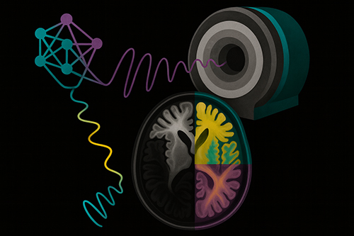
A novel, dictionary-free, multi-contrast MRI method for microscopic imaging
This project aims to develop a novel quantitative imaging technique for comprehensive in vitro and in vivo tissue characterisation on the microscopic scale. The technology innovated in the project could revolutionise microscopic imaging techniques by breaking through the sub-millimetre image resolution bottleneck of current magnetic resonance imaging (MRI) methods. This project expects to generate new knowledge in the emerging field of biological imaging and to deliver an integrated imaging platform for mapping various tissue microscopic components at the cellular level. Successful outcomes have the potential for commercialisation and will accelerate a range of fundamental science and engineering studies requiring imaging techniques.
Project Lead: Dr Hongfu Sun
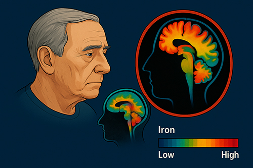
Disambiguating Parkinson’s disease from disorders with mimicking symptoms using ultra-high-field (7 Tesla) multi-modal MRI
Differentiating the clinical diagnosis of Parkinson’s disease (PD) from disorders with which it is often confused is challenging even for experienced neurologists, particularly in the early disease stages when patients first present. Misdiagnosing patients with parkinsonism can lead to incorrect treatment decisions and significant distress for patients and families because of the considerable prognosis variability in the different underlying pathologies. This project aims to develop technological innovations toward significant clinical applications for diagnosing Parkinson’s disease from disorders with mimicking symptoms. In addition to improved diagnosis, these discoveries will offer potential mechanistic insights into the diseases by accurately mapping regional brain iron content.
Project Lead: Dr Hongfu Sun and Prof Peter Nestor
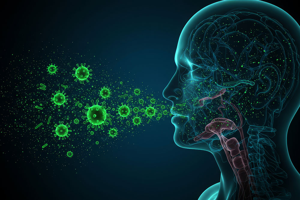
Respiratory Virus Identifying Breathalyser
This project aims to develop a breathalyser capable of rapidly identifying respiratory viruses by detecting virus-specific extracellular vesicles (EVs) in exhaled breath. The team will use an advanced aptamer-based diagnostic technology, where aptamers (single-stranded DNA oligonucleotides) are developed to bind specifically to EVs released by virus-infected airway cells. These EVs carry viral proteins, making them ideal biomarkers for early and non-invasive detection. The aptamers will be selected using sequential evolution of ligands by exponential enrichment (SELEX) and immobilised on gold electrodes to enable sensitive detection via electrochemical impedance spectroscopy. Ultimately, the goal is to create a scalable, multiplex-capable diagnostic device that can detect multiple respiratory viruses simultaneously, with commercialisation discussions already underway. This technology will not only support timely diagnosis and treatment at the individual level but also enable large-scale community surveillance to monitor outbreaks and emerging threats.
Project lead: Dr Renee Goreham and Prof Nathan Bartlett
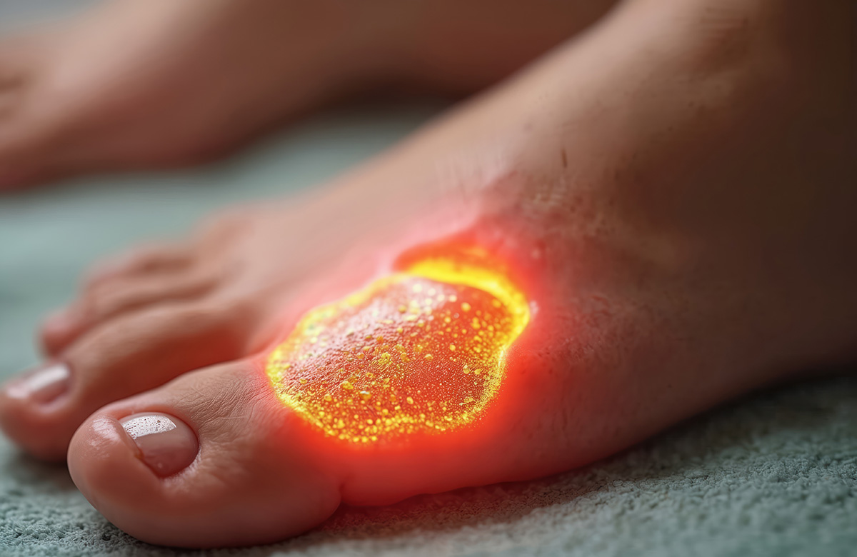
Multispectral fluorescence imaging for detecting bacterial infections and monitoring wound progression in clinical environments
In collaboration with Whiteley, Genesys Design and University of Western Sydney, and federally funded through a CRC-P project we aim to design a portable fluorescent imaging device capable of imaging biofilm in chronic wounds.
Project lead: Prof Sarah Johnson
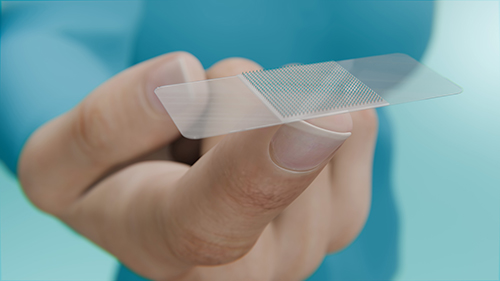
Fabrication of Microneedles for Drug Delivery and Biosensing
This project aims to develop a simple, cost-effective method for fabricating microneedles, with a focus on applications in painless drug delivery and biosensing. The microneedles are made from biocompatible and biodegradable materials and are less than 1 mm in length, allowing them to painlessly penetrate the outer skin layer for drug delivery or biomarker detection for diagnostic purposes.
Project lead: A/Prof Yuen Yong and Dr Van Dau (Griffith University)
Collaborators
We partner with:
- Clinical centres: John Hunter Hospital, Liverpool Hospital, Mater Hospital Brisbane
- Industry leaders: Whiteley and Genesys Design
- National and international research institutes, including the University of Western Sydney, Griffith University, HMRI Infection Research Program.
The University of Newcastle acknowledges the traditional custodians of the lands within our footprint areas: Awabakal, Darkinjung, Biripai, Worimi, Wonnarua, and Eora Nations. We also pay respect to the wisdom of our Elders past and present.