Centre Facilities
The Global Centre for Research and Training in Radiation Oncology state-of-the-art facilities include a Linear Accelerator in a university setting, in what is a world-first.
The facilities provide a safe environment for students to gain clinical skills in an academic setting. They develop confidence in their abilities by undertaking hands-on training before entering the clinical world. Access to the Global Centre's radiation oncology equipment and software creates confident, life-ready graduates, ultimately advancing the delivery of cancer care and the patient experience.
Our laboratories incorporate 'Outreach Cameras'; with the ability to deliver training and engage in research projects accessible anywhere in the world. Students and clinical partners have access to the most experienced educators and world-class medical technology and equipment.
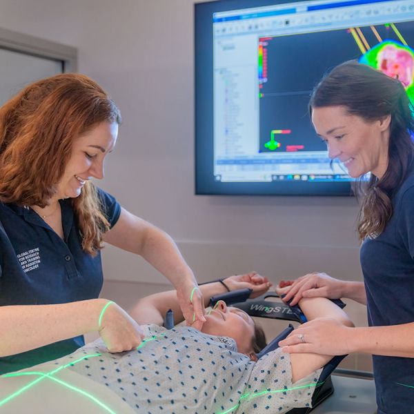
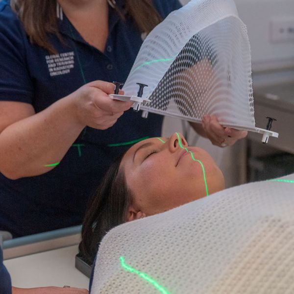
Simulation Laboratory
Features: 8 simulation bays, clinical grade immobilisation and tattooing stations.
The Simulation Laboratory allows students to perform clinical skills like creating immobilisation devices for patient treatments and patient positioning using surface anatomy. Students practice skills surrounding the mapping of a patient for custom Radiation Therapy treatment. The Simulation Laboratory has equipment required for all cancer sites, which are used to immobilise and create comfort for patients to deliver safe and optimal treatment.
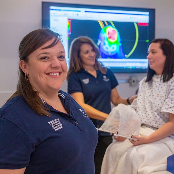
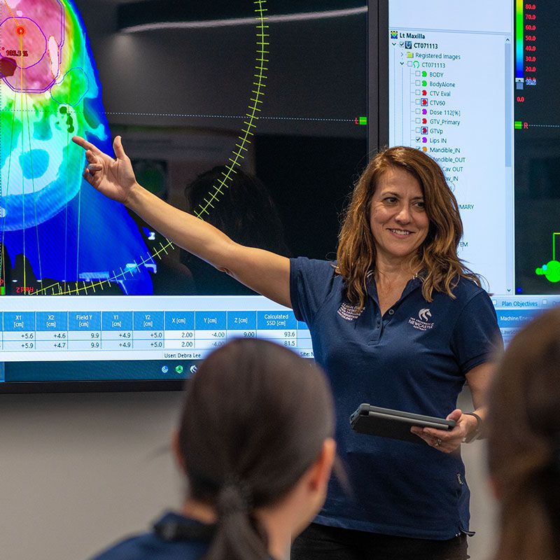
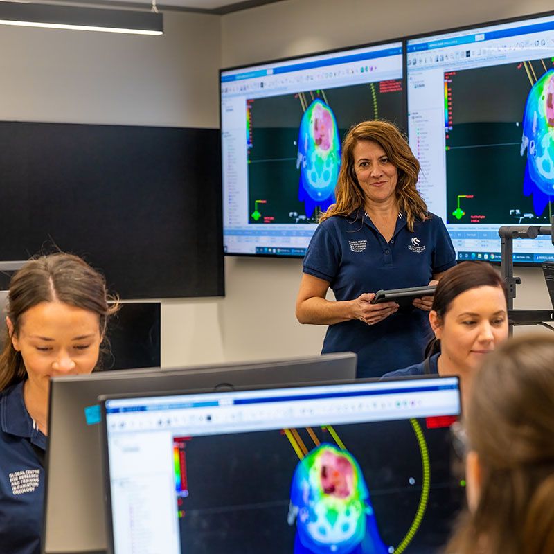
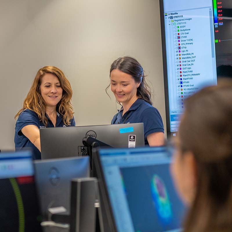
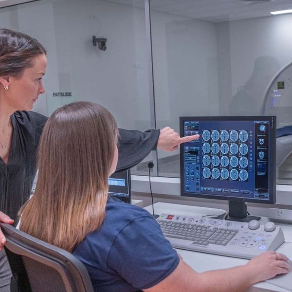
Planning Laboratory
Features: 36 PCs with Eclipse and Monaco treatment planning software systems as well as ProKnow software.
The Planning Laboratory is used in the preparation of ‘Radiation Therapy Plans’. Radiation Therapy is ‘planned’ for each patient as a customised treatment on specialised computer software. Students have access to cutting-edge, clinically-relevant planning software and are trained in the leading Radiation Therapy planning techniques used in clinical settings.
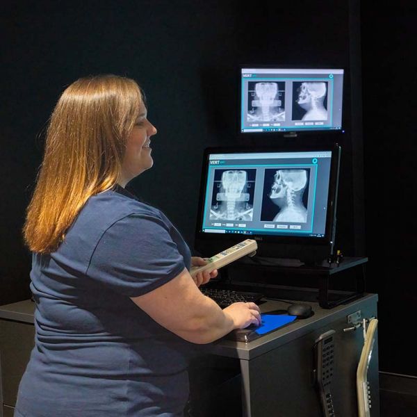
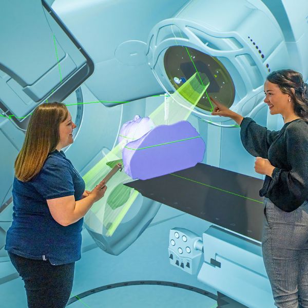
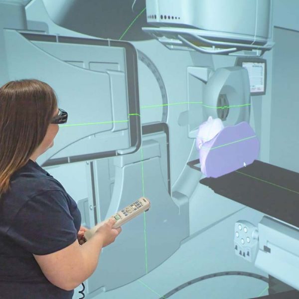
Virtual Laboratory
Features: 3-D learning tool, with clinical DICOM data, image guided radiation therapy (IGRT) practice function and positioning and anatomy training.
The VERT system uses sophisticated 3-D technology to simulate real-life human anatomy and cancerous tumours from anonymised medical images/datasets. This technology gives students hands-on practice with patient treatment setups, reinforcing anatomy knowledge and anatomical positioning terms.
It has a function to practice IGRT – the use of imaging during radiation therapy to improve the precision and accuracy of treatment delivery. By overlaying the image taken for the planning process with daily treatment setup images, treatment is delivered with millimetre precision.
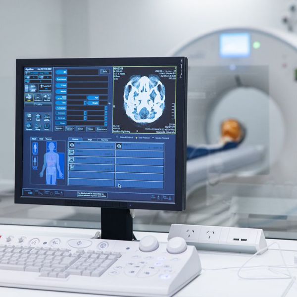
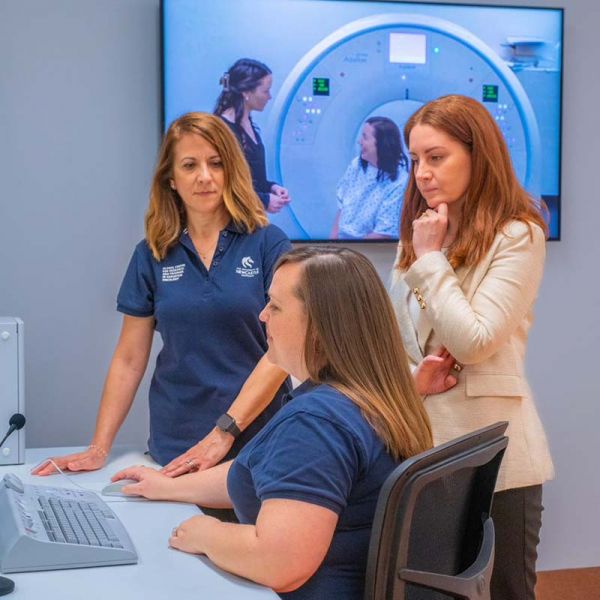
Lightening Aquilion Computed Tomography
Features: fully functional CT capabilities with clinical grade immobilisation.
The CT laboratory is equipped with a fully functioning 'Canon Lightening Aquilion' CT scanner. It includes:
- A console area, with CT protocols for radiation therapy CT data acquisition
- CCTV and microphone capabilities, for 'patient' monitoring and communication
- Phantoms for CT acquisition practice
- Clinical grade immobilisation devices
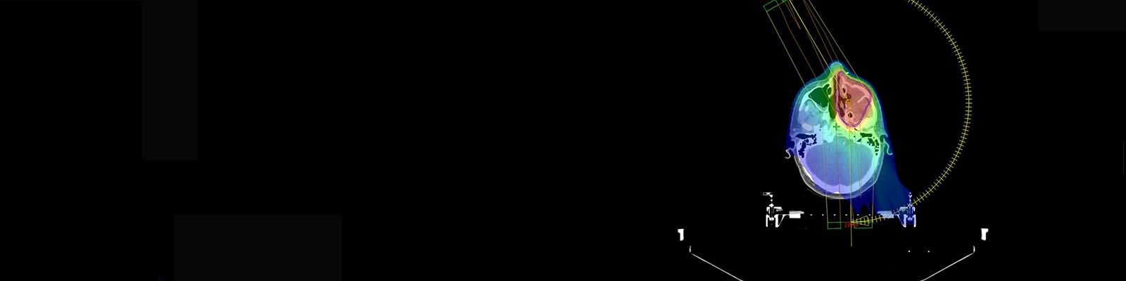
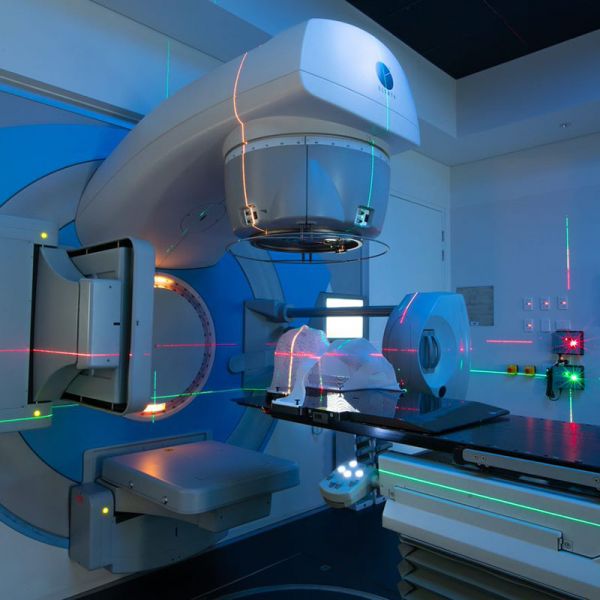
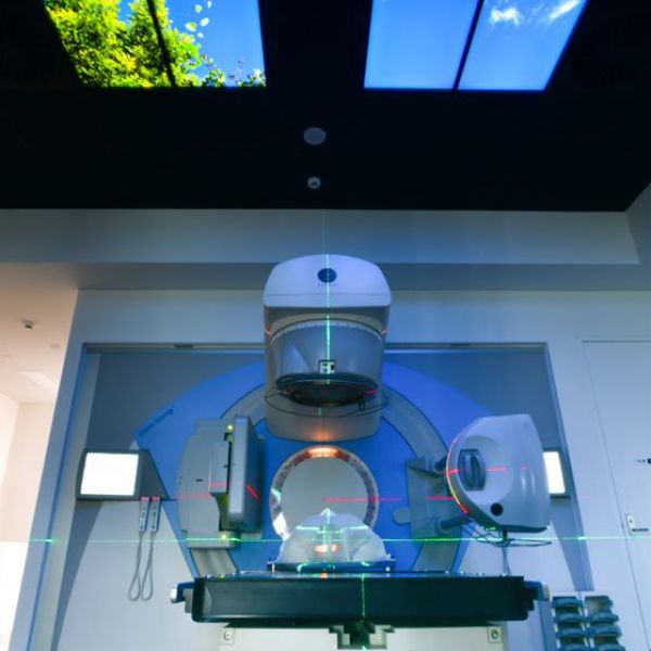
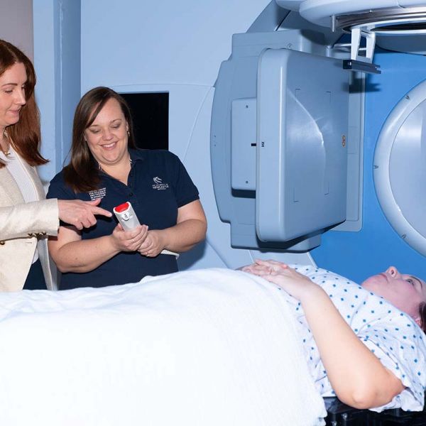
Elekta Linear Accelerator Bunker
Features: A Linear Accelerator (fully functional however not radiation producing).
The Elekta Linear Accelerator (Versa HDTM) is located within a purpose-built bunker, designed to simulate a clinical setting. It includes:
- A console area, lasers and simulated imaging capabilities
- ‘C-RAD’ cameras for Surface-Guided Radiation Therapy (SGRT)
- ‘Active Breathing Coordinator’ (ABC) gating system
- Clinical grade immobilisation devices
- The bunker also has a 'bell' mounted on the wall, to resemble that of clinical sites, where patients celebrate their final Radiation Therapy treatment. When the bell is rung, staff, patients and loved ones join in on the very triumphant and emotional moment and cheer on the patient when they finish treatment.
The University of Newcastle acknowledges the traditional custodians of the lands within our footprint areas: Awabakal, Darkinjung, Biripai, Worimi, Wonnarua, and Eora Nations. We also pay respect to the wisdom of our Elders past and present.