Researcher Highlights
Rethinking radiation therapy
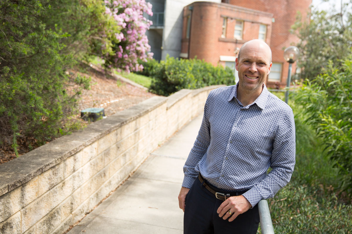
Conjoint Professor Peter Greer
Combining expertise in the fields of physics, medicine, and IT, Peter Greer and his team are marrying research and practice.
Working quietly behind the scenes in every radiation oncology centre, are a group of scientists who manage the extremely intricate processes involved in the delivery of radiation oncology.
Medical physicists work to implement and develop imaging techniques to delineate tumours, dose calculation algorithms to calculate complex doses, and intensity modulated radiation delivery to deliver highly tailored dose distributions of radiation therapy.
Peter currently holds a full-time research position leading a medical physicis research group at the Calvary Mater Newcastle Hospital and University of Newcastle.
His work aims to improve the treatment of cancer patients with radiation therapy and enable high quality effective treatments.
Much awarded, Peter is recognised as a world leader in the field of radiation dosimetry with flat-panel imaging devices, and has twice taken the role of Scientific Director for the major international conference in radiotherapy treatment imaging.
MRI planning and delivery
Along with surgery, radiation oncology is a frontline treatment for cancers.
Delivered either internally of externally depending on the tumour site, radiotherapy technology, which uses high-energy x-ray beams to sterilise DNA in tumors, continues to improve.
Not only do medical physicists ensure all aspects of treatment are precisely calculated and delivered, but they also drive the development of improved technology and techniques through research.
Investigating MRIs as a potential game changer in radiation oncology, Peter and his team have been working in collaboration with CSIRO Biomedical Imaging Research Group at the Australian E-Health Research Centre since 2005.
“MRI is fantastic for delineating the tumours and the normal tissues,” Peter says.
“But we are currently still reliant on CT scans for calculating the dose.”
A true pioneer in this area, Peter has developed the first atlas-based deformable image registration method to map realistic electron densities to MRI scans for dose calculations.
“If we can avoid the CT scanning, and just plan directly with the MRI, that would cost less for the health system, it's better for the patient, and it would be more accurate treatment as well.”
Having proven effective for treatment dosage calculations in retrospective studies, Peter and his team are now beginning to implement the improved MRI-only system for prospective prostrate tumour patients in the clinic.
Watchdog
Collaborating with Cancer Care Manitoba and Memorial Sloan Kettering, Peter and his team are also developing Watchdog, a real-time verification imaging system.
Conventional radiotherapy has been reliant on the accuracy of pre-emptive dosage calculations, with no way of verifying delivery during treatment.
“Major errors are very rare in Australia,” Peter notes.
“But minor errors are more frequent, and underdosing or overdosing could compromise treatment.”
A prestigious grant from The American Society for Radiation Oncology (ASTRO) recognises the massive positive impacts a real-time verification system could have on patient safety.
“We are now verifying the delivery using the imaging system in a movie mode,” Peter explains
“The real-time measurement allows for immediate checking, and if something is not right you just stop, and it really doesn't require any extra effort.”
The National Health and Medical Research Council (NHMRC) has also contributed to the research, funding the team to develop a successful prototype into a system that can be used by any radiation oncology centre.
Remote auditing
Also based at the Calvary Mater Hospital is The Trans-Tasman Radiation Oncology Group (TROG), a global leader in radiotherapy research.
With TROG research threatened by ever increasing costs associated with sending physicists to remote multicenter research sites to ensure treatment uniformity, Peter came up with a solution.
The Virtual EPID Standard Phantom Audit (VESPA) uses imaging systems to record EPID images then sent electronically to TROG for remote dosing analysis and audit.
So successful have trials of this system been that Peter and his team are now assisting TROG’s UK equivalent - The Cancer Research UK and UCL Cancer Trials Centre - to trial this method of remote auditing.
Possibilities and Paths
Peter was drawn to medical physics because he believed this path would include ever changing challenges, and the opportunity to see research implemented.
“Everything changes so quickly, there is always new technology, so we have to be constantly thinking and upgrading our knowledge,” Peter says.
He credits his extensive experience on the clinical coalface as his inspiration for ideas regarding optimal treatments.
“Sometimes things are obvious and lots of people are looking at them,” he says.
“But sometimes it is just a matter of working in an area for a while, and then seeing the possibilities and paths.”
Asked if he is ever overwhelmed by the necessity of his clinical work, Peter responds immediately.
“There is always hope, and I am grateful my work is making a positive difference.”

The University of Newcastle acknowledges the traditional custodians of the lands within our footprint areas: Awabakal, Darkinjung, Biripai, Worimi, Wonnarua, and Eora Nations. We also pay respect to the wisdom of our Elders past and present.