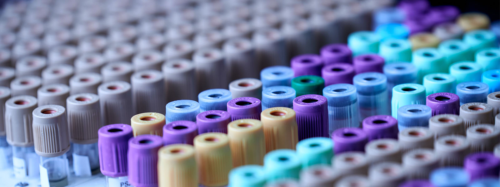
Central Analytical Facilities
Our Central Analytical Facilities team gives researchers, students and external partners access to specialised scientific equipment. Our instruments are supported by expert staff and are centrally maintained.
Our impact
Analytical Mass Spectrometry
Chemical analyses for elemental (atomic) and low-weight molecules. Application areas are wide-ranging and include clinical and biological research as well as the environmental, geochemical, forensic, pharmaceutical, food and beverage, and petrochemical industries.
Read more
Biological Mass Spectrometry
Helps detect, identify, and quantify proteins, peptides, small molecules and metabolites. Supports biomedical, chemical, biological and environmental research.
Read more
Flow Cytometry
Detects and measures the physical and chemical characteristics of cellular and particle parameters at the single cell level.
Read more
Advanced Confocal Microscopy
Captures super-resolution 2D and 3D images from tissue sections, including live and fixed cells. Supports biological and biomedical research.
Read more
Scanning Electron Microscopy
Produces sharp, high-resolution images that give researchers topographical, morphological and compositional information. Supports medical, biological, material sciences and engineering.
Read more
Transmission Electron Microscopy
A multi-task platform capable of analysing ultra-thin sections or particles. Supports advanced material manufacturing, next generation energy storage solutions, environmental remediation, industrial catalysts, molecule and drug delivery design, and biomedical research.
Read more
X-Ray Technology
X-Ray Diffraction is a powerful technique used in material and life sciences, physics, chemistry, pharmaceuticals and manufacturing. It uses focused X-ray beams to determine chemical compositions, characterise crystalline structures, identify minerals and more.
Read more
Equipment access and pricing
The University of Newcastle acknowledges the traditional custodians of the lands within our footprint areas: Awabakal, Darkinjung, Biripai, Worimi, Wonnarua, and Eora Nations. We also pay respect to the wisdom of our Elders past and present.