
Professor Hubert Hondermarck
Professor
School of Biomedical Sciences and Pharmacy (Medical Biochemistry)
- Email:hubert.hondermarck@newcastle.edu.au
- Phone:(02) 4921 8830
Career Summary
Biography
Hubert Hondermarck is a biochemist specialised in Cancer Neuroscience. He obtained a PhD in neurobiochemistry at the University of Lille, France (1990) and was a post-doctoral researcher at the University of California Irvine (1990-1993) where he investigated the molecular mechanisms of neuronal cell differentiation in Professor Ralph A.Bradshaw laboratory. He then created a research unit of the French Institute of Health and Medical Research (U908, INSERM) dedicated to the study of growth factor signaling and functional proteomics in cancer. In 2011, he relocated to the University of Newcastle Australia, to start a new program on Cancer Neuroscience, to investigate the role of the nervous system, neurotrophic growth factors and neuromolecules in cancer.
Research Expertise: Cancer Neuroscience
The Hondermarck research group investigates the crosstalk between nerves and cancer cells, and its impact on tumour growth and metastasis. Until recently neurons were thought not to be involved in cancer. However, recent evidence including from our laboratory, have introduced the new paradigm that nerves actually promote tumour initiation and progression. Denervation can suppress both the development of the primary tumour and the outburst of metastases. The objective of this research is to identify the molecular mediators of the crosstalk between nerves and cancer cells and develop them as innovative clinical biomarkers and therapeutic targets in oncology. The methodologies include the analysis of human tumour samples, cell cultures, proteomics and mass spectrometry analysis. We work in collaboration with neurobiologists, pathologists, clinicians and private companies to translate the results of our research into practical outcomes in oncology.
%2FPic%20research.jpg)
Figure: Nerve-cancer cell crosstalk. Nerves infiltrate the tumor microenvironment and stimulate cancer cell growth and metastasis through the secretion of neurotransmitters (such as catecholamines, acetylcholine and neuropeptides) initiating signaling pathways for growth and invasion in cancer cells after binding to neurotransmitter receptors (NTRs). Conversely, nerve infiltration in the tumor is mediated through the liberation of neurotrophic growth factors (such as NGF) by cancer cells, resulting in neuron outgrowth (axonogenesis or neo-neurogenesis), as well as autocrine stimulation of cancer cells via the stimulation of corresponding receptor tyrosine kinases (RTKs). This reciprocal interaction fuels tumor development and also impacts the microenvironment, as the liberated neurotransmitters and growth factors can also act on endothelial and immune cells, then contributing to tumor inflammation and neo-angiogenesis. Cancer-induced pain can also be a consequence of tumor innervation. PLCγ, phospholipase C gamma; cAMP, cyclic adenosine monophosphate; STAT, signal transducer activator of transcription; PKC, protein kinase C; MAPK, mitogen-activated protein kinases. From our review Jobling et al. Cancer Res. (2015).
Full publication list: https://pubmed.ncbi.nlm.nih.gov/?term=hondermarck&sort=date
Representative recent publications:
Magnon C, Hondermarck H. The neural addiction of cancer. Nat Rev Cancer. 2023 May;23(5):317-334.
Hondermarck H, Jiang CC. Time to Introduce Nerve Density in Cancer Histopathological Assessment. Clin Cancer Res. 2023 Apr 28:CCR-23-0736.
Jiang CC, Marsland M, Wang Y, Dowdell A, Eden E, Gao F, Faulkner S, Jobling P, Li X, Liu L, He Z, Hondermarck H. Tumor innervation is triggered by endoplasmic reticulum stress. Oncogene. 2022 Jan;41(4):586-599. doi: 10.1038/s41388-021-02108-6. Epub 2021 Nov 16. PMID: 34785777.
Li X, Liu H, Dun MD, Faulkner S, Liu X, Jiang CC, Hondermarck H. Proteome and secretome analysis of pancreatic cancer cells. Proteomics. 2022 Jul;22(13-14):e2100320.
Delahunt B, Steigler A, Atkinson C, Christie D, Duchesne G, Egevad L, Joseph D, Kenwright DN, Matthews J, Murray JD, Oldmeadow C, Samaratunga H, Spry NA, Thunders MC, Hondermarck H, Denham JW. Percentage grade 4 tumour predicts outcome for prostate adenocarcinoma in needle biopsies from patients with advanced disease: 10-year data from the TROG 03.04 RADAR trial. Pathology. 2022 Feb;54(1):49-54.
Monje M, Borniger JC, D'Silva NJ, Deneen B, Dirks PB, Fattahi F, Frenette PS, Garzia L, Gutmann DH, Hanahan D, Hervey-Jumper SL, Hondermarck H, Hurov JB, Kepecs A, Knox SM, Lloyd AC, Magnon C, Saloman JL, Segal RA, Sloan EK, Sun X, Taylor MD, Tracey KJ, Trotman LC, Tuveson DA, Wang TC, White RA, Winkler F. Roadmap for the Emerging Field of Cancer Neuroscience. Cell. 2020 Apr 16;181(2):219-222.
Faulkner S, Griffin N, Rowe CW, Jobling P, Lombard JM, Oliveira SM, Walker MM, Hondermarck H. Nerve growth factor and its receptor tyrosine kinase TrkA are overexpressed in cervical squamous cell carcinoma. FASEB Bioadv. 2020 Jun 30;2(7):398-408.
Gao F, Griffin N, Faulkner S, Li X, King SJ, Jobling P, Denham JW, Jiang CC, Hondermarck H. The Membrane Protein Sortilin Can Be Targeted to Inhibit Pancreatic Cancer Cell Invasion. Am J Pathol. 2020 Sep;190(9):1931-1942.
Griffin N, Rowe CW, Gao F, Jobling P, Wills V, Walker MM, Faulkner S, Hondermarck H. Clinicopathological Significance of Nerves in Esophageal Cancer. Am J Pathol. 2020 Sep;190(9):1921-1930.
March B, Faulkner S, Jobling P, Steigler A, Blatt A, Denham J, Hondermarck H. Tumour innervation and neurosignalling in prostate cancer. Nat Rev Urol. 2020 Feb;17(2):119-130.
Delahunt B, Murray JD, Steigler A, Atkinson C, Christie D, Duchesne G, Egevad L, Joseph D, Matthews J, Oldmeadow C, Samaratunga H, Spry NA, Srigley JR, Hondermarck H, Denham JW. Perineural invasion by prostate adenocarcinoma in needle biopsies predicts bone metastasis: Ten year data from the TROG 03.04 RADAR Trial. Histopathology. 2020 Apr 13.
Rowe CW, Dill T, Griffin N, Jobling P, Faulkner S, Paul JW, King S, Smith R, Hondermarck H. Innervation of papillary thyroid cancer and its association with extra-thyroidal invasion. Sci Rep. 2020 Jan 30;10(1):1539.
Liu H, Li X, Dun MD, Faulkner S, Jiang CC, Hondermarck H. Cold Shock Domain Containing E1 (CSDE1) Protein is Overexpressed and Can be Targeted to Inhibit Invasiveness in Pancreatic Cancer Cells. Proteomics. 2020 May;20(10):e1900331.
Faulkner S, Jobling P, March B, Jiang CC, Hondermarck H.Tumor Neurobiology and the War of Nerves in Cancer. Cancer Discov. 2019, 9(6):702-710.
Editorial boards
Hubert Hondermarck is a member of the Editorial Board of the following Journals: Molecular and Cellular Proteomics (ASBMB), FASEB Bioadvances (Wiley), Proteomics (Wiley), Proteomics Clinical Applications (Wiley), Open Cancer Journal (Bentham), Frontiers in Endocrinology (Frontiers Media).
Teaching
Biochemistry, cancer biology, proteomics, bioinformatics.
Course coordinator: HUBS3302 Bioinformatic and functional genomics. HUBS3409. Projects in Biomedical Sciences.
Administrative Activities
Head of the Discipline of Medical Biochemistry (Faculty of Health and Medicine).
Qualifications
- PhD (Life Sciences & Health), University of Lille - France
Keywords
- Cancer Neuroscience
- Proteomics
Languages
- English (Fluent)
- French (Mother)
Fields of Research
| Code | Description | Percentage |
|---|---|---|
| 321109 | Predictive and prognostic markers | 50 |
| 321199 | Oncology and carcinogenesis not elsewhere classified | 50 |
Professional Experience
UON Appointment
| Title | Organisation / Department |
|---|---|
| Professor | University of Newcastle School of Biomedical Sciences and Pharmacy Australia |
Publications
For publications that are currently unpublished or in-press, details are shown in italics.
Book (1 outputs)
| Year | Citation | Altmetrics | Link | |||||
|---|---|---|---|---|---|---|---|---|
| 1997 |
Bradshaw RA, Hondermarck H, Nerve growth factor receptors (1997)
|
|||||||
Chapter (5 outputs)
| Year | Citation | Altmetrics | Link | ||||||||
|---|---|---|---|---|---|---|---|---|---|---|---|
| 2022 |
Hondermarck H, Bradshaw RA, 'Cell Communication An Overview', Encyclopedia of Cell Biology: Volume 1-6, Second Edition 1-3 (2022) Cell communication is the driving force that enables the development and functioning of all multicellular organisms. From reproduction and embryogenesis to cellular and tissular h... [more] Cell communication is the driving force that enables the development and functioning of all multicellular organisms. From reproduction and embryogenesis to cellular and tissular homeostasis in the adult, the control of metabolism and general physiology, as well as the dysregulations in pathological processes, cell communication is germane to all aspects of eukaryotic life. This section covers cell signaling in a holistic way by detailing the principal extracellular and intracellular mechanisms underlying cell communication.
|
||||||||||
| 2022 |
Skerrett-Byrne D, Jiang CC, Nixon B, Hondermarck H, 'Transcriptomics', Encyclopedia of Cell Biology (Second Edition), Elsevier, Amsterdam, Netherlands 363-371 (2022)
|
||||||||||
| 2016 |
Milward EA, Shahandeh A, Heidari M, Johnstone DM, Daneshi N, Hondermarck H, 'Transcriptomics', Encyclopedia of Cell Biology, Elsevier, The Netherland 160-165 (2016)
|
||||||||||
| 2015 |
Hondermarck H, Demont Y, Bradshaw RA, 'The TrK receptor family', Receptor Tyrosine Kinases: Family and Subfamilies, Springer, New York 777-820 (2015) [B1]
|
Nova | |||||||||
| Show 2 more chapters | |||||||||||
Journal article (161 outputs)
| Year | Citation | Altmetrics | Link | ||||||||
|---|---|---|---|---|---|---|---|---|---|---|---|
| 2024 |
Boilly B, Hondermarck H, Aguado MT, 'Nerves and availability of mesodermal cells are essential for the function of the segment addition zone (SAZ) during segment regeneration in polychaete annelids.', Dev Genes Evol, (2024) [C1]
|
||||||||||
| 2024 |
Amit M, Anastasaki C, Dantzer R, Demir IE, Deneen B, Dixon KO, et al., 'Next Directions in the Neuroscience of Cancers Arising outside the CNS.', Cancer Discov, 14 669-673 (2024)
|
||||||||||
| 2023 |
Hondermarck H, Jiang CC, 'Time to Introduce Nerve Density in Cancer Histopathologic Assessment', CLINICAL CANCER RESEARCH, 29 2342-2344 (2023)
|
Nova | |||||||||
| 2023 |
Li RQ, Zhao XH, Zhu Q, Liu T, Hondermarck H, Thorne RF, et al., 'Exploring neurotransmitters and their receptors for breast cancer prevention and treatment.', Theranostics, 13 1109-1129 (2023) [C1]
|
Nova | |||||||||
| 2023 |
Magnon C, Hondermarck H, 'The neural addiction of cancer.', Nat Rev Cancer, 23 317-334 (2023) [C1]
|
Nova | |||||||||
| 2023 |
Dowdell A, Marsland M, Faulkner S, Gedye C, Lynam J, Griffin CP, et al., 'Targeting XBP1 mRNA splicing sensitizes glioblastoma to chemotherapy', FASEB BIOADVANCES, 5 211-220 (2023) [C1]
|
Nova | |||||||||
| 2023 |
Winter M, Nait Eldjoudi A, Guette C, Hondermarck H, Bourette RP, Fovez Q, et al., 'Mitochondrial adaptation decreases drug sensitivity of persistent triple negative breast cancer cells surviving combinatory and sequential chemotherapy.', Neoplasia, 46 100949 (2023) [C1]
|
Nova | |||||||||
| 2023 |
Marsland M, Dowdell A, Faulkner S, Gedye C, Lynam J, Griffin CP, et al., 'The Membrane Protein Sortilin Is a Potential Biomarker and Target for Glioblastoma', Cancers, 15 2514-2514 [C1]
|
Nova | |||||||||
| 2023 |
Marsland M, Dowdell A, Faulkner S, Jobling P, Rush RA, Gedye C, et al., 'ProNGF Expression and Targeting in Glioblastoma Multiforme.', Int J Mol Sci, 24 (2023) [C1]
|
Nova | |||||||||
| 2022 |
Boilly B, Hondermarck H, Boilly-Marer Y, 'Neural regulation of body polarities in nereid worm regeneration', FASEB BioAdvances, 4 22-28 (2022) [C1] Nerve dependence in regeneration has been established more than 200¿years ago but the mechanisms by which nerves are necessary to regeneration remain to be fully elucidated. Aside... [more] Nerve dependence in regeneration has been established more than 200¿years ago but the mechanisms by which nerves are necessary to regeneration remain to be fully elucidated. Aside from their direct impact in stimulating cellular growth, nerves also have a role on the establishment of body polarities (antero-posterior and dorso-ventral patterns) and this has been particularly well studied in nereid annelid worms. Nereids can regenerate appendages (parapodia) and the tail (body segments). In both parapodia and tail regeneration, the presence of the nerve cord is necessary to the establishment of body polarities. In this review, we will detail the experimental procedures which have been conducted in nereids to elucidate the role of the nerve cord in the establishment of the antero-posterior and dorso-ventral polarities. Most of the studies reported here were published several decades ago and based on anatomical and histological analyses; this review should constitute a knowledgebase and an inspiration for needed modern-time explorations at the molecular levels to elucidate the impact of the nervous system in the acquisition of body polarities.
|
Nova | |||||||||
| 2022 |
Griffin CP, Paul CL, Alexander KL, Walker MM, Hondermarck H, Lynam J, 'Postmortem brain donations vs premortem surgical resections for glioblastoma research: viewing the matter as a whole.', Neurooncol Adv, 4 vdab168 (2022) [C1]
|
Nova | |||||||||
| 2022 |
Li D, Hu LN, Zheng SM, La T, Wei LY, Zhang XJ, et al., 'High nerve density in breast cancer is associated with poor patient outcome', FASEB BioAdvances, 4 391-401 (2022) [C1] Active crosstalk between the nervous system and breast cancer cells has been experimentally demonstrated in vitro and in animal models. However, low frequencies of peripheral nerv... [more] Active crosstalk between the nervous system and breast cancer cells has been experimentally demonstrated in vitro and in animal models. However, low frequencies of peripheral nerve presence in human breast cancers reported in previous studies (~30% of cases) potentially negate a major role of the nervous system in breast cancer development and progression. This study aimed to clarify the incidence of nerves within human breast cancers and to delineate associations with clinicopathological features. Immunohistochemical staining was conducted in formalin-fixed paraffin-embedded breast cancer tissue sections using antibodies against the pan-neuronal markers protein gene product 9.5 and growth-associated protein 43, and the sympathetic nerve-specific marker tyrosine hydroxylase. Nerve trunks and isolated nerve fibers were quantitated. The chi-squared test was used to determine the associations between nerve counts and clinicopathological parameters. The log-rank test was used to compare differences in patient progression-free survival (PFS) and overall survival (OS). The overall frequency of peripheral nerves in breast cancers was 85%, a markedly higher proportion than reported previously. Of note, most nerves present in breast cancers were of the sympathetic origin. While high density of nerve trunks or isolated nerve fibers was associated with poor PFS and OS of patients, high nerve trunk density appeared also to predict poor patient PFS independently of lymph node metastasis. Innervation of breast cancers is a common event correlated with poor patient outcomes. These findings support the notion that the nervous system plays an active role in breast cancer pathogenesis.
|
Nova | |||||||||
| 2022 |
Ferdoushi A, Jamaluddin MFB, Li X, Pundavela J, Faulkner S, Hondermarck H, 'Secretome analysis of human schwann cells derived from malignant peripheral nerve sheath tumor', PROTEOMICS, 22 (2022) [C1]
|
Nova | |||||||||
| 2022 |
Delahunt B, Steigler A, Atkinson C, Christie D, Duchesne G, Egevad L, et al., 'Percentage grade 4 tumour predicts outcome for prostate adenocarcinoma in needle biopsies from patients with advanced disease: 10-year data from the TROG 03.04 RADAR trial', Pathology, 54 49-54 (2022) [C1] Previous reports have shown that quantification of high tumour grade is of prognostic significance for patients with prostate cancer. In particular, percent Gleason pattern 4 (GP4... [more] Previous reports have shown that quantification of high tumour grade is of prognostic significance for patients with prostate cancer. In particular, percent Gleason pattern 4 (GP4) has been shown to predict outcome in several studies, although conflicting results have also been reported. A major issue with these studies is that they rely on surrogate markers of outcome rather than patient survival. We have investigated the prognostic predictive value of quantifying GP4 in a series of prostatic biopsies containing Gleason score 3+4=7 and 4+3=7 tumours. It was found that the length of GP4 tumour determined from the measurement of all biopsy cores from a single patient, percent GP4 present and absolute GP4 were all significantly associated with distant progression of tumour, all-cause mortality and cancer-specific mortality over a 10-year follow-up period. Assessment of the relative prognostic significance showed that these parameters outperformed division of cases according to Gleason score (3+4=7 versus 4+3=7). International Society of Urological Pathology (ISUP) Grade Groups currently divide these tumours, according to Gleason grading guidelines, into grade 2 (3+4=7) and grade 3 (4+3=7). Our results indicate that this simple classification results in the loss of important prognostic information. In view of this we would recommend that ISUP Grade Groups 2 and 3 be amalgamated as grade 2 tumour with the percentage of GP4 carcinoma being appended to the final grade, e.g., 3+4=7 carcinoma with 40% pattern 4 tumour would be classified as ISUP Grade Group 2 (40%).
|
Nova | |||||||||
| 2022 |
Jiang CC, Marsland M, Wang Y, Dowdell A, Eden E, Gao F, et al., 'Tumor innervation is triggered by endoplasmic reticulum stress', ONCOGENE, 41 586-599 (2022) [C1]
|
Nova | |||||||||
| 2022 |
Li X, Liu H, Dun MD, Faulkner S, Liu X, Jiang CC, Hondermarck H, 'Proteome and secretome analysis of pancreatic cancer cells', PROTEOMICS, 22 (2022) [C1]
|
Nova | |||||||||
| 2022 |
Marsland M, Dowdell A, Jiang CC, Wilmott JS, Scolyer RA, Zhang XD, et al., 'Expression of NGF/proNGF and Their Receptors TrkA, p75(NTR) and Sortilin in Melanoma', INTERNATIONAL JOURNAL OF MOLECULAR SCIENCES, 23 (2022) [C1]
|
Nova | |||||||||
| 2021 |
March B, Lockhart KR, Faulkner S, Smolny M, Rush R, Hondermarck H, 'ELISA-based quantification of neurotrophic growth factors in urine from prostate cancer patients', FASEB BioAdvances, 3 888-896 (2021) [C1] Non-invasive procedures are needed for prostate cancer management, and urine represents a potential source of new biomarkers with translational value. Recent evidence has shown th... [more] Non-invasive procedures are needed for prostate cancer management, and urine represents a potential source of new biomarkers with translational value. Recent evidence has shown that the growth of new nerves in the tumor microenvironment is essential to prostate cancer progression. Neurotrophic growth factors are expressed by prostate cancer cells and contribute to prostate tumor innervation, but their presence in urine is unclear. In the present study, we have assayed the concentration of neurotrophic factors in the urine of prostate cancer patients. Urine was collected from a prospective cohort of 45 men with prostate cancer versus 30 men without cancer and enzyme-linked immunosorbent assay was used to quantify nerve growth factor (NGF) and its precursor proNGF, brain-derived neurotrophic factor (BDNF) and proBDNF, neurotrophin-3, neurotrophin-4/5, and glia-derived neurotrophic growth factor. The results show that neurotrophic factors are detectable in various concentrations in both cancer and healthy urine, but no significant difference was found. Also, no association was observed between neurotrophic factor concentrations and prostate cancer grade. This study is the first quantification of neurotrophins in urine, and although no significant differences were observed between prostate cancer patients versus those without prostate cancer, or between prostate cancers of various grades, the potential value of neurotrophins for prostate cancer diagnosis and prognosis warrants further investigations in larger patient cohorts.
|
Nova | |||||||||
| 2021 |
Ferdoushi A, Griffin N, Marsland M, Xu X, Faulkner S, Gao F, et al., 'Tumor innervation and clinical outcome in pancreatic cancer', SCIENTIFIC REPORTS, 11 (2021) [C1]
|
Nova | |||||||||
| 2021 |
Griffin N, Gao F, Jobling P, Oldmeadow C, Wills V, Walker MM, et al., 'The neurotrophic tyrosine kinase receptor 1 (TrkA) is overexpressed in oesophageal squamous cell carcinoma', Pathology, 53 470-477 (2021) [C1] Nerve growth factor (NGF) and its receptors, the neurotrophic receptor tyrosine kinase 1 (NTRK1/TrkA) and the common neurotrophin receptor (NGFR/p75NTR), are increasingly implicat... [more] Nerve growth factor (NGF) and its receptors, the neurotrophic receptor tyrosine kinase 1 (NTRK1/TrkA) and the common neurotrophin receptor (NGFR/p75NTR), are increasingly implicated in cancer progression, but their clinicopathological significance in oesophageal cancer is unclear. In this study, the expression of NGF, NTRK1 and NGFR were analysed by immunohistochemistry in a cohort of 303 oesophageal cancers versus 137 normal adjacent oesophageal tissues. Immunostaining was digitally quantified and compared to clinicopathological parameters. NGF and NGFR staining were found in epithelial cells and at similar levels between oesophageal cancers and normal oesophageal tissue. NGFR staining was slightly increased with grade (p=0.0389). Interestingly, NTRK1 staining was markedly higher in oesophageal squamous cell carcinoma (OR 2.31, 95%CI 1.13¿4.38, p<0.0001) and significantly lower in adenocarcinoma (OR 0.50, 95%CI 0.44¿0.63, p<0.0001) compared to normal oesophageal tissue. In addition, NTRK1 staining was decreased in grade 2 and grade 3 (OR 0.51, 95%CI 0.21¿1.40, p<0.0001) compared to grade 1, suggesting a preferential involvement of this receptor in the more differentiated forms of oesophageal carcinomas. Together, these data point to NTRK1 as a biomarker and a candidate therapeutic target in oesophageal squamous cell carcinoma.
|
Nova | |||||||||
| 2021 |
Ding J, Zhang R, Li H, Ji Q, Cheng X, Thorne RF, et al., 'ASIC1 and ASIC3 mediate cellular senescence of human nucleus pulposus mesenchymal stem cells during intervertebral disc degeneration.', Aging, 13 10703-10723 (2021) [C1]
|
Nova | |||||||||
| 2021 |
Hondermarck H, Huang PS, Wagner JA, 'The nervous system: Orchestra conductor in cancer, regeneration, inflammation and immunity', FASEB BioAdvances, 3 944-952 (2021) [C1] Although the role of nerves in stimulating cellular growth and dissemination has long been described in tissue regeneration studies, until recently a similar trophic role of nerve... [more] Although the role of nerves in stimulating cellular growth and dissemination has long been described in tissue regeneration studies, until recently a similar trophic role of nerves in disease was not well recognized. However, recent studies in oncology have demonstrated that the growth and dissemination of cancers also requires the infiltration of nerves in the tumor microenvironment. Nerves generate various neurosignaling pathways, which orchestrate cancer initiation, progression, and metastases. Similarly, nerves are increasingly implicated for their regulatory functions in immunity and inflammation. This orchestrator role of nerves in cellular and molecular interactions during regeneration, cancer, immunity, and inflammation offers new possibilities for targeting or enhancing neurosignaling in human health and diseases.
|
Nova | |||||||||
| 2020 |
Griffin N, Rowe CW, Gao F, Jobling P, Wills V, Walker MM, et al., 'Clinicopathological Significance of Nerves in Esophageal Cancer', American Journal of Pathology, 190 1921-1930 (2020) [C1] Nerves are emerging promoters of cancer progression, but the innervation of esophageal cancer and its clinicopathologic significance remain unclear. In this study, nerves were ana... [more] Nerves are emerging promoters of cancer progression, but the innervation of esophageal cancer and its clinicopathologic significance remain unclear. In this study, nerves were analyzed by immunohistochemistry in a cohort of 260 esophageal cancers, including 40 matched lymph node metastases and 137 normal adjacent esophageal tissues. Nerves were detected in 38% of esophageal cancers and were more associated with squamous cell carcinomas (P = 0.04). The surrounding or invasion of nerves by cancer cells (perineural invasion) was detected in 12% of esophageal cancers and was associated with reduced survival (P = 0.04). Nerves were found to express the following receptors for nerve growth factor (NGF): neurotrophic receptor tyrosine kinase 1 and nerve growth factor receptor. An association was suggested between high production of NGF by cancer cells and the presence of nerves (P = 0.02). In vitro, NGF production in esophageal cancer cells was shown by Western blot, and esophageal cancer cells were able to induce neurite outgrowth in the PC12 neuronal cells. The neurotrophic activity of esophageal cancer cells was inhibited by anti-NGF blocking antibodies. Together, these data suggest that innervation is a feature in esophageal cancers that may be driven by cancer cell¿released NGF.
|
Nova | |||||||||
| 2020 |
Faulkner S, Griffin N, Rowe CW, Jobling P, Lombard JM, Oliveira SM, et al., 'Nerve growth factor and its receptor tyrosine kinase TrkA are overexpressed in cervical squamous cell carcinoma.', FASEB bioAdvances, 2 398-408 (2020) [C1]
|
Nova | |||||||||
| 2020 |
Delahunt B, Murray JD, Steigler A, Atkinson C, Christie D, Duchesne G, et al., 'Perineural invasion by prostate adenocarcinoma in needle biopsies predicts bone metastasis: Ten year data from the TROG 03.04 RADAR Trial', Histopathology, 77 284-292 (2020) [C1] Aims: Perineural invasion (PNI) by prostatic adenocarcinoma is debated as a prognostic parameter. This study investigates the prognostic predictive value of PNI in a series of pat... [more] Aims: Perineural invasion (PNI) by prostatic adenocarcinoma is debated as a prognostic parameter. This study investigates the prognostic predictive value of PNI in a series of patients with locally advanced prostate cancer treated with radiotherapy and androgen deprivation using 10¿years outcome data from the TROG 03.04 RADAR trial. Methods: Diagnostic prostate biopsies from 976 patients were reviewed and the presence of PNI noted. Patients were followed for 10¿years according to the trial protocol or until death. The primary endpoint for the study was time to bone metastasis. Secondary endpoints included time to soft tissue metastasis, transition to castration resistance, prostate cancer-specific mortality and all-cause mortality. Results: PNI was detected in 449 cases (46%), with 234 cases (24%) having PNI in more than one core. The presence of PNI was significantly associated with higher ISUP grade, clinical T staging category, National Comprehensive Cancer Network risk group, and percent positive biopsy cores. The cumulative probability of bone metastases according to PNI status was significant over the 10¿years follow-up interval of the study (log-rank test P¿<¿0.0001). PNI was associated with all endpoints on univariable analysis. After adjusting for baseline clinicopathological and treatment factors, bone metastasis was the only endpoint in which PNI retained its prognostic significance (hazard ratio 1.42, 95% confidence interval 1.05¿1.92, P¿=¿0.021). Conclusions: The association between PNI and the development of bone metastases supports the inclusion of this parameter as a component of the routine histology report. Further this association suggests that evaluation of PNI may assist in selecting those patients who should be monitored more closely during follow-up.
|
Nova | |||||||||
| 2020 |
Ferdoushi A, Li X, Griffin N, Faulkner S, Jamaluddin MFB, Gao F, et al., 'Schwann Cell Stimulation of Pancreatic Cancer Cells: A Proteomic Analysis', Frontiers in Oncology, 10 (2020) [C1]
|
Nova | |||||||||
| 2020 |
Gao F, Griffin N, Faulkner S, Li X, King SJ, Jobling P, et al., 'The Membrane Protein Sortilin Can Be Targeted to Inhibit Pancreatic Cancer Cell Invasion.', The American journal of pathology, 190 (2020) [C1]
|
Nova | |||||||||
| 2020 |
Netherton J, Ogle RA, Hetherington L, Villaverde AISB, Hondermarck H, Baker MA, 'Proteomic analysis reveals that topoisomerase 2A is associated with defective sperm head morphology', Molecular and Cellular Proteomics, 19 444-455 (2020) [C1]
|
Nova | |||||||||
| 2020 |
Griffin N, Marsland M, Roselli S, Oldmeadow C, Attia J, Walker MM, et al., 'The receptor tyrosine kinase trka is increased and targetable in HER2-positive breast cancer', Biomolecules, 10 1-13 (2020) [C1] The tyrosine kinase receptor A (NTRK1/TrkA) is increasingly regarded as a therapeutic target in oncology. In breast cancer, TrkA contributes to metastasis but the clinicopathologi... [more] The tyrosine kinase receptor A (NTRK1/TrkA) is increasingly regarded as a therapeutic target in oncology. In breast cancer, TrkA contributes to metastasis but the clinicopathological significance remains unclear. In this study, TrkA expression was assessed via immunohistochemistry of 158 invasive ductal carcinomas (IDC), 158 invasive lobular carcinomas (ILC) and 50 ductal carcinomas in situ (DCIS). TrkA was expressed in cancer epithelial and myoepithelial cells, with higher levels of TrkA positively associated with IDC (39% of cases) (p < 0.0001). Interestingly, TrkA was significantly increased in tumours expressing the human epidermal growth factor receptor-2 (HER2), with expression in 49% of HER2-positive compared to 25% of HER2-negative tumours (p = 0.0027). A panel of breast cancer cells were used to confirm TrkA protein expression, demonstrating higher levels of TrkA (total and phosphorylated) in HER2-positive cell lines. Functional investigations using four different HER2-positive breast cancer cell lines indicated that the Trk tyrosine kinase inhibitor GNF-5837 reduced cell viability, through decreased phospho-TrkA (Tyr490) and downstream AKT (Ser473) activation, but did not display synergy with Herceptin. Overall, these data highlight a relationship between the tyrosine kinase receptors TrkA and HER2 and suggest the potential of TrkA as a novel or adjunct target for HER2-positive breast tumours.
|
Nova | |||||||||
| 2020 |
Liu H, Li X, Dun MD, Faulkner S, Jiang CC, Hondermarck H, 'Cold Shock Domain Containing E1 (CSDE1) Protein is Overexpressed and Can be Targeted to Inhibit Invasiveness in Pancreatic Cancer Cells', PROTEOMICS, 20 (2020) [C1]
|
Nova | |||||||||
| 2020 |
Rowe CW, Dill T, Griffin N, Jobling P, Faulkner S, Paul JW, et al., 'Innervation of papillary thyroid cancer and its association with extra-thyroidal invasion', Scientific Reports, 10 (2020) [C1]
|
Nova | |||||||||
| 2020 |
Dun MD, Mannan A, Rigby CJ, Butler S, Toop HD, Beck D, et al., 'Shwachman Bodian Diamond syndrome (SBDS) protein is a direct inhibitor of protein phosphatase 2A (PP2A) activity and overexpressed in acute myeloid leukaemia', Leukemia, 34 3393-3397 (2020) [C1]
|
Nova | |||||||||
| 2020 |
March B, Faulkner S, Jobling P, Steigler A, Blatt A, Denham J, Hondermarck H, 'Tumour innervation and neurosignalling in prostate cancer', Nature Reviews Urology, 17 119-130 (2020) [C1] Prostate cancer progression has been shown to be dependent on the development of autonomic nerves into the tumour microenvironment. Sympathetic nerves activate adrenergic neurosig... [more] Prostate cancer progression has been shown to be dependent on the development of autonomic nerves into the tumour microenvironment. Sympathetic nerves activate adrenergic neurosignalling that is necessary in early stages of tumour progression and for initiating an angiogenic switch, whereas parasympathetic nerves activate cholinergic neurosignalling resulting in tumour dissemination and metastasis. The innervation of prostate cancer seems to be initiated by neurotrophic growth factors, such as the precursor to nerve growth factor secreted by tumour cells, and the contribution of brain-derived neural progenitor cells has also been reported. Current experimental, epidemiological and clinical evidence shows the stimulatory effect of tumour innervation and neurosignalling in prostate cancer. Using nerves and neurosignalling could have value in the management of prostate cancer by predicting aggressive disease, treating localized disease through denervation and relieving cancer-associated pain in bone metastases.
|
Nova | |||||||||
| 2020 |
Hondermarck H, Bartlett NW, Nurcombe V, 'The role of growth factor receptors in viral infections: An opportunity for drug repurposing against emerging viral diseases such as COVID-19?', FASEB bioAdvances, 2 296-303 (2020) [C1]
|
Nova | |||||||||
| 2019 |
Ferdoushi A, Li X, Bin Jamaluddin MF, Hondermarck H, 'Proteomic Profile of Human Schwann Cells', PROTEOMICS, 20 (2019) [C1]
|
Nova | |||||||||
| 2019 |
Jezequel P, Kerdraon O, Hondermarck H, Guerin-Charbonnel C, Lasla H, Gouraud W, et al., 'Identification of three subtypes of triple-negative breast cancer with potential therapeutic implications', BREAST CANCER RESEARCH, 21 (2019) [C1]
|
Nova | |||||||||
| 2019 |
Bradshaw RA, Hondermarck H, Rodriguez H, 'Cancer Proteomics and the Elusive Diagnostic Biomarkers', PROTEOMICS, 19 (2019) [C1]
|
Nova | |||||||||
| 2019 |
Nurcombe V, Ling L, Hondermarck H, Cool SM, Smith RAA, 'Bringing Heparan Sulfate Glycomics Together with Proteomics for the Design of Novel Therapeutics: A Historical Perspective', Proteomics, 19 (2019) [C1] Increasing knowledge of how peptides bind saccharides, and of how saccharides bind peptides, is starting to revolutionize understanding of cell-extracellular matrix relationships.... [more] Increasing knowledge of how peptides bind saccharides, and of how saccharides bind peptides, is starting to revolutionize understanding of cell-extracellular matrix relationships. Here, a historical perspective is taken of the relationship between heparan sulfate glycosaminoglycans and how they interact with peptide growth factors in order to both drive and modulate signaling through the appropriate cognate receptors. Such knowledge is guiding the preparation of targeted sugar mimetics that will impact the treatment of many different kinds of diseases, including cancer.
|
Nova | |||||||||
| 2019 |
Rowe CW, Faulkner S, Paul JW, Tolosa JM, Gedye C, Bendinelli C, et al., 'The precursor for nerve growth factor (proNGF) is not a serum or biopsy-rinse biomarker for thyroid cancer diagnosis.', BMC endocrine disorders, 19 128 (2019) [C1]
|
Nova | |||||||||
| 2019 |
Rowe CW, Dill T, Faulkner S, Gedye C, Paul JW, Tolosa JM, et al., 'The precursor for nerve growth factor (ProNGF) in thyroid cancer lymph node metastases: Correlation with primary tumour and pathological variables', International Journal of Molecular Sciences, 20 1-13 (2019) [C1]
|
Nova | |||||||||
| 2019 |
Duchatel RJ, Jackson ER, Alvaro F, Nixon B, Hondermarck H, Dun MD, 'Signal Transduction in Diffuse Intrinsic Pontine Glioma', PROTEOMICS, 19 (2019) [C1]
|
Nova | |||||||||
| 2019 |
Faulkner S, Jobling P, March B, Jiang CC, Hondermarck H, 'Tumor neurobiology and the war of nerves in cancer', Cancer Discovery, 9 702-710 (2019) [C1] Nerves are emerging regulators of cancer progression. Cancer cells induce the outgrowth of nerves in the tumor microenvironment through the release of neu-rotrophic factors, and i... [more] Nerves are emerging regulators of cancer progression. Cancer cells induce the outgrowth of nerves in the tumor microenvironment through the release of neu-rotrophic factors, and in return nerves liberate neurotransmitters that activate cancer growth and dissemination. Although sympathetic nerves drive tumor angiogenesis via the liberation of noradrena-line, sensory and parasympathetic nerves stimulate cancer stem cells. Interestingly, recent evidence indicates that parasympathetic nerves can eventually inhibit tumor progression, suggesting a yin¿yang type of regulation of cancer by nerves. From a broader perspective, the question of a higher level of control of cancer development by the central nervous system should be raised. Significance: Nerves are emerging regulators of cancer initiation, progression, and metastasis. Here, we review the evidence to date and explore the basic and clinical ramifications of these findings.
|
Nova | |||||||||
| 2019 |
Ahmed AF, de Bock CE, Sontag E, Hondermarck H, Lincz LF, Thorne RF, 'FAT1 cadherin controls neuritogenesis during NTera2 cell differentiation', Biochemical and Biophysical Research Communications, 514 625-631 (2019) [C1]
|
Nova | |||||||||
| 2019 |
Gholizadeh N, Greer PB, Simpson J, Denham J, Lau P, Dowling J, et al., 'Characterization of prostate cancer using diffusion tensor imaging: a new perspective', European Journal of Radiology, 110 112-120 (2019) [C1]
|
Nova | |||||||||
| 2018 |
Almazi JG, Pockney P, Gedye C, Smith ND, Hondermarck H, Verrills NM, Dun MD, 'Cell-Free DNA Blood Collection Tubes Are Appropriate for Clinical Proteomics: A Demonstration in Colorectal Cancer.', Proteomics. Clinical applications, 12 e1700121 (2018) [C1]
|
Nova | |||||||||
| 2018 |
Gao F, Griffin N, Faulkner S, Rowe CW, Williams L, Roselli S, et al., 'The neurotrophic tyrosine kinase receptor TrkA and its ligand NGF are increased in squamous cell carcinomas of the lung', SCIENTIFIC REPORTS, 8 (2018) [C1]
|
Nova | |||||||||
| 2018 |
Griffin N, Faulkner S, Jobling P, Hondermarck H, 'Targeting neurotrophin signaling in cancer: The renaissance', Pharmacological Research, 135 12-17 (2018) [C1] Nerve outgrowth in the tumor microenvironment (tumor neurogenesis) has recently been shown to be essential for cancer progression and the concept of nerve dependence is emerging i... [more] Nerve outgrowth in the tumor microenvironment (tumor neurogenesis) has recently been shown to be essential for cancer progression and the concept of nerve dependence is emerging in oncology. Neurotrophins such as nerve growth factor (NGF) have long been identified as drivers of neurogenesis during development and regeneration, but intriguingly they were also known to be expressed in human tumors where they can stimulate cancer cell growth. Recent findings have unraveled that NGF released by cancer cells is also a driver of tumor neurogenesis, via the stimulation of NGF receptors on nerve endings. In return, nerves infiltrated in the tumor microenvironment secrete neurotransmitters, which can stimulate both the growth of tumor cells and angiogenesis. This neurotrophic role of NGF in cancer is likely to be relevant to a large variety of human malignancies, as well as other neurotrophins, and may have ramifications in cancer pain. Therefore, pharmacological interventions against neurotrophin signaling have the potential not only to target cancer cells directly, but also to inhibit neurogenesis and its stimulatory impact on cancer progression and pain.
|
Nova | |||||||||
| 2018 |
Li X, Dun MD, Faulkner S, Hondermarck H, 'Neuroproteins in Cancer: Assumed Bystanders Become Culprits', PROTEOMICS, 18 (2018) [C1]
|
Nova | |||||||||
| 2018 |
Faulkner S, Jobling P, Rowe CW, Rodrigues Oliveira SM, Roselli S, Thorne RF, et al., 'Neurotrophin Receptors TrkA, p75 Neurotrophin receptors are emerging targets in oncology, but their clinicopathologic significance in thyroid cancer is unclear. In this study, the neurotrophin tyrosine receptor k... [more] Neurotrophin receptors are emerging targets in oncology, but their clinicopathologic significance in thyroid cancer is unclear. In this study, the neurotrophin tyrosine receptor kinase TrkA (also called NTRK1), the common neurotrophin receptor p75NTR, and the proneurotrophin receptor sortilin were analyzed with immunohistochemistry in a cohort of thyroid cancers (n = 128) and compared with adenomas and normal thyroid tissues (n = 62). TrkA was detected in 20% of thyroid cancers, compared with none of the benign samples (P = 0.0007). TrkA expression was independent of histologic subtypes but associated with lymph node metastasis (P = 0.0148), suggesting the involvement of TrkA in tumor invasiveness. Nerves in the tumor microenvironment were positive for TrkA. p75NTR was overexpressed in anaplastic thyroid cancers compared with papillary and follicular subtypes (P < 0.0001). Sortilin was overexpressed in thyroid cancers compared with benign thyroid tissues (P < 0.0001). Neurotrophin receptor expression was confirmed in a panel of thyroid cancer cell lines at the mRNA and protein levels. Functional investigations using the anaplastic thyroid cancer cell line CAL-62 found that siRNA against TrkA, p75NTR, and sortilin decreased cell survival and cell migration through decreased SRC and ERK activation. Together, these data reveal TrkA, p75NTR, and sortilin as potential therapeutic targets in thyroid cancer.
|
Nova | |||||||||
| 2018 |
Sahoo SS, Zhang XD, Hondermarck H, Tanwar PS, 'The Emerging Role of the Microenvironment in Endometrial Cancer', CANCERS, 10 (2018) [C1]
|
Nova | |||||||||
| 2018 |
Jamaluddin MFB, Ko YA, Kumar M, Brown Y, Bajwa P, Nagendra PB, et al., 'Proteomic profiling of human uterine fibroids reveals upregulation of the extracellular matrix protein periostin', Endocrinology, 159 1106-1118 (2018) [C1] The central characteristic of uterine fibroids is excessive deposition of extracellular matrix (ECM), which contributes to fibroid growth and bulk-type symptoms. Despite this, ver... [more] The central characteristic of uterine fibroids is excessive deposition of extracellular matrix (ECM), which contributes to fibroid growth and bulk-type symptoms. Despite this, very little is known about patterns of ECM protein expression in fibroids and whether these are influenced by the most common genetic anomalies, which relate to MED12. We performed extensive genetic and proteomic analyses of clinically annotated fibroids and adjacent normal myometrium to identify the composition and expression patterns of ECM proteins in MED12 mutation-positive and mutation-negative uterine fibroids. Genetic sequencing of tissue samples revealed MED12 alterations in 39 of 65 fibroids (60%) from 14 patients. Using isobaric tagged-based quantitative mass spectrometry on three selected patients (n = 9 fibroids), we observed a common set of upregulated (.1.5-fold) and downregulated (,0.66-fold) proteins in small, medium, and large fibroid samples of annotated MED12 status. These two sets of upregulated and downregulated proteins were the same in all patients, regardless of variations in fibroid size and MED12 status. We then focused on one of the significant upregulated ECM proteins and confirmed the differential expression of periostin using western blotting and immunohistochemical analysis. Our study defined the proteome of uterine fibroids and identified that increased ECM protein expression, in particular periostin, is a hallmark of uterine fibroids regardless of MED12 mutation status. This study sets the foundation for further investigations to analyze the mechanisms regulating ECM overexpression and the functional role of upregulated ECM proteins in leiomyogenesis.
|
Nova | |||||||||
| 2017 |
Boilly B, Faulkner S, Jobling P, Hondermarck H, 'Nerve Dependence: From Regeneration to Cancer', Cancer Cell, 31 342-354 (2017) [C1] Nerve dependence has long been described in animal regeneration, where the outgrowth of axons is necessary to the reconstitution of lost body parts and tissue remodeling in variou... [more] Nerve dependence has long been described in animal regeneration, where the outgrowth of axons is necessary to the reconstitution of lost body parts and tissue remodeling in various species. Recent discoveries have demonstrated that denervation can suppress tumor growth and metastasis, pointing to nerve dependence in cancer. Regeneration and cancer share similarities in regard to the stimulatory role of nerves, and there are indications that the stem cell compartment is a preferred target of innervation. Thus, the neurobiology of cancer is an emerging discipline that opens new perspectives in oncology.
|
Nova | |||||||||
| 2017 |
Guo ST, Guo XY, Wang J, Wang CY, Yang RH, Wang FH, et al., 'MicroRNA-645 is an oncogenic regulator in colon cancer', ONCOGENESIS, 6 (2017) [C1]
|
Nova | |||||||||
| 2017 |
Rougemont B, Bontemps Gallo S, Ayciriex S, Carrière R, Hondermarck H, Lacroix JM, et al., 'Scout-MRM: Multiplexed Targeted Mass Spectrometry-Based Assay without Retention Time Scheduling Exemplified by Dickeya dadantii Proteomic Analysis during Plant Infection', Analytical Chemistry, 89 1421-1426 (2017) [C1]
|
Nova | |||||||||
| 2017 |
Rutledge A, Jobling P, Walker MM, Denham JW, Hondermarck H, 'Spinal Cord Injuries and Nerve Dependence in Prostate Cancer', Trends in Cancer, 3 812-815 (2017) [C1] Nerves are emerging as drivers of tumorigenesis, as demonstrated in the mouse where denervation suppresses prostate cancer; however, clinical evidence is needed. Patients with spi... [more] Nerves are emerging as drivers of tumorigenesis, as demonstrated in the mouse where denervation suppresses prostate cancer; however, clinical evidence is needed. Patients with spinal cord injuries (SCIs) resulting in functional denervation of the prostate have a lower incidence of prostate cancer. This may constitute a clinical evidence for nerve dependence in human prostate tumorigenesis.
|
Nova | |||||||||
| 2017 |
Shargh VH, Hondermarck H, Liang M, 'Gelatin-albumin hybrid nanoparticles as matrix metalloproteinases-degradable delivery systems for breast cancer therapy', Nanomedicine, 12 977-989 (2017) [C1] Aim: To develop matrix metalloproteinase-responsive gelatin-albumin hybrid nanoparticles encapsulating a selective tropomyosin receptor kinase A (TrkA) inhibitor GNF-5837 (Gel-Alb... [more] Aim: To develop matrix metalloproteinase-responsive gelatin-albumin hybrid nanoparticles encapsulating a selective tropomyosin receptor kinase A (TrkA) inhibitor GNF-5837 (Gel-Alb-GNF HNPs) and to demonstrate their anticancer effects in breast cancer. Methods: Gel-Alb-GNF HNPs were prepared using a pH-controlled complexation process from cationic gelatin, dextran sulfate and albumin-bound GNF-5837. The anticancer activities of Gel-Alb-GNF HNPs were tested in a panel of subtype-specific breast cancer cell lines. Results: Gel-Alb-GNF HNPs (~130 nm) displayed excellent stability and matrix metalloproteinase-triggered drug release. Compared with GNF-5837 alone, Gel-Alb-GNF HNPs not only significantly enhanced the antiproliferative and anti-invasive effects but also restored the apoptosis of cancer cells. Conclusion: Gel-Alb-GNF HNPs may be adaptable for stand-alone therapies or used in combination with traditional chemotherapies for breast cancer treatment.
|
Nova | |||||||||
| 2017 |
de Bock CE, Hughes MR, Snyder K, Alley S, Sadeqzadeh E, Dun MD, et al., 'Protein interaction screening identifies SH3RF1 as a new regulator of FAT1 protein levels', FEBS LETTERS, 591 667-678 (2017) [C1]
|
Nova | |||||||||
| 2016 |
Faulkner S, Roselli S, Demont Y, Pundavela J, Choquet G, Leissner P, et al., 'ProNGF is a potential diagnostic biomarker for thyroid cancer', Oncotarget, 7 28488-28497 (2016) [C1] The precursor for nerve growth factor (proNGF) is expressed in some cancers but its clinicopathological significance is unclear. The present study aimed to define the clinicopatho... [more] The precursor for nerve growth factor (proNGF) is expressed in some cancers but its clinicopathological significance is unclear. The present study aimed to define the clinicopathological significance of proNGF in thyroid cancer. ProNGF expression was analysed by immunohistochemistry in two cohorts of cancer versus benign tumors (adenoma) and normal thyroid tissues. In the first cohort (40 thyroid cancers, 40 thyroid adenomas and 80 normal thyroid tissues), proNGF was found overexpressed in cancers compared to adenomas and normal samples (p<0.0001). The area under the receiver-operating characteristic (ROC) curve was 0.84 (95% CI 0.75-0.93, p<0.0001) for cancers versus adenomas, and 0.99 (95% CI 0.98-1.00, p<0.0001) for cancers versus normal tissues. ProNGF overexpression was confirmed in a second cohort (127 cancers of various histological types and 55 normal thyroid tissues) and using a different antibody (p<0.0001). ProNGF staining intensity was highest in papillary carcinomas compared to other histological types (p<0.0001) and there was no significant association with age, gender, tumor size, stage and lymph node status. In conclusion, proNGF is increased in thyroid cancer and should be considered as a new potential diagnostic biomarker.
|
Nova | |||||||||
| 2016 |
Hetherington L, Schneider EK, DeKretser D, Muller CH, Hondermarck H, Velkov T, et al., 'Deficiency in outer dense fiber 1 is a marker and potential driver of idiopathic male infertility', Molecular and Cellular Proteomics, 15 3685-3693 (2016) [C1] Globally, ~1 in 15 men of reproductive age are infertile, yet the precise mechanisms underlying their gamete failure are unknown. Although a semen analysis is performed to determi... [more] Globally, ~1 in 15 men of reproductive age are infertile, yet the precise mechanisms underlying their gamete failure are unknown. Although a semen analysis is performed to determine fertilizing potential, the diagnostic suitability of this analysis has been questioned in several reports, as many men, classified as infertile according to their semen analysis, subsequently turn out to be fertile. Herein, we have used a quantitative (phospho)-proteomic analysis, using enrichment on titanium dioxide followed by ion-trap mass spectrometry (LC-MS/MS), to compare the semen of infertile versus fertile males. One protein, namely outer dense fiber 1 (ODF1), was dramatically reduced in infertile males. Using specific antibodies, we then screened the gametes of a cohort of suspected infertile men and demonstrated a reduction in the amount of ODF1 compared with fertile controls. Stress treatment of sperm deficient in ODF1 caused the head to decapitate, suggesting why these gametes fail to initiate fertilization. Interestingly, electron micrographs of ODF1-deficient spermatozoa revealed an abnormal connecting piece, indicating several developmental defects with both the implantation plate and the thin laminated fibers. In some cases, the implantation plate appeared to be reduced in size or was overburdened by granular material near the connecting piece. Hence, a strong reduction ODF1 is a marker of idiopathic male infertility and a potential driver of this condition.
|
Nova | |||||||||
| 2016 |
Shargh VH, Hondermarck H, Liang M, 'Antibody-targeted biodegradable nanoparticles for cancer therapy', Nanomedicine, 11 63-79 (2016) [C1] The use of nanotechnology has great potentials to revolutionize the future cancer diagnosis and therapy. In this context, various nanoparticles (NPs) have been developed for targe... [more] The use of nanotechnology has great potentials to revolutionize the future cancer diagnosis and therapy. In this context, various nanoparticles (NPs) have been developed for targeted delivery of diagnostic/therapeutic agents to the tumor sites, which thus result in greater efficacy and much less side effects. The targeting property of NPs is often achieved by functionalizing their surface with tumor-specific ligands, such as antibodies, peptides, small molecules and oligonucleotides. In this review, we will discuss recent progress in the multifunctional design of antibody-targeted NPs with a special focus on liposomal, polymeric and protein-based delivery systems.
|
Nova | |||||||||
| 2016 |
Guo ST, Chi MN, Yang RH, Guo XY, Zan LK, Wang CY, et al., 'INPP4B is an oncogenic regulator in human colon cancer', Oncogene, 35 3049-3061 (2016) [C1] Inositol polyphosphate 4-phosphatase type II (INPP4B) negatively regulates phosphatidylinositol 3-kinase signaling and is a tumor suppressor in some types of cancers. However, we ... [more] Inositol polyphosphate 4-phosphatase type II (INPP4B) negatively regulates phosphatidylinositol 3-kinase signaling and is a tumor suppressor in some types of cancers. However, we have found that it is frequently upregulated in human colon cancer cells. Here we show that silencing of INPP4B blocks activation of Akt and serum-and glucocorticoid-regulated kinase 3 (SGK3), inhibits colon cancer cell proliferation and retards colon cancer xenograft growth. Conversely, overexpression of INPP4B increases proliferation and triggers anchorage-independent growth of normal colon epithelial cells. Moreover, we demonstrate that the effect of INPP4B on Akt and SGK3 is associated with inactivation of phosphate and tensin homolog through its protein phosphatase activity and that the increase in INPP4B is due to Ets-1-mediated transcriptional upregulation in colon cancer cells. Collectively, these results suggest that INPP4B may function as an oncogenic driver in colon cancer, with potential implications for targeting INPP4B as a novel approach to treat this disease.
|
Nova | |||||||||
| 2016 |
Shargh VH, Hondermarck H, Liang M, 'Albumin hybrid nanoparticles loaded with tyrosine kinase A inhibitor GNF-5837 for targeted inhibition of breast cancer cell growth and invasion.', Int J Pharm, 515 527-534 (2016) [C1]
|
Nova | |||||||||
| 2015 |
Dun MD, Chalkley RJ, Faulkner S, Keene S, Avery-Kiejda KA, Scott RJ, et al., 'Proteotranscriptomic profiling of 231-BR breast cancer cells: Identification of potential biomarkers and therapeutic targets for brain metastasis', Molecular and Cellular Proteomics, 14 2316-2330 (2015) [C1] Brain metastases are a devastating consequence of cancer and currently there are no specific biomarkers or therapeutic targets for risk prediction, diagnosis, and treatment. Here ... [more] Brain metastases are a devastating consequence of cancer and currently there are no specific biomarkers or therapeutic targets for risk prediction, diagnosis, and treatment. Here the proteome of the brain metastatic breast cancer cell line 231-BR has been compared with that of the parental cell line MDA-MB-231, which is also metastatic but has no organ selectivity. Using SILAC and nanoLC-MS/MS, 1957 proteins were identified in reciprocal labeling experiments and 1584 were quantified in the two cell lines. A total of 152 proteins were confidently determined to be up- or down-regulated by more than twofold in 231-BR. Of note, 112/152 proteins were decreased as compared with only 40/152 that were increased, suggesting that down-regulation of specific proteins is an important part of the mechanism underlying the ability of breast cancer cells to metastasize to the brain. When matched against transcriptomic data, 43% of individual protein changes were associated with corresponding changes in mRNA, indicating that the transcript level is a limited predictor of protein level. In addition, differential miRNA analyses showed that most miRNA changes in 231-BR were up- (36/45) as compared with down-regulations (9/45). Pathway analysis revealed that proteome changes were mostly related to cell signaling and cell cycle, metabolism and extracellular matrix remodeling. The major protein changes in 231-BR were confirmed by parallel reaction monitoring mass spectrometry and consisted in increases (by more than fivefold) in the matrix metalloproteinase-1, ephrin-B1, stomatin, myc target-1, and decreases (by more than 10-fold) in transglutaminase-2, the S100 calcium-binding protein A4, and L-plastin. The clinicopathological significance of these major proteomic changes to predict the occurrence of brain metastases, and their potential value as therapeutic targets, warrants further investigation.
|
Nova | |||||||||
| 2015 |
Roselli S, Pundavela J, Demont Y, Faulkner S, Keene S, Attia J, et al., 'Sortilin is associated with breast cancer aggressiveness and contributes to tumor cell adhesion and invasion', Oncotarget, 6 10473-10486 (2015) [C1] The neuronal membrane protein sortilin has been reported in a few cancer cell lines, but its expression and impact in human tumors is unclear. In this study, sortilin was analyzed... [more] The neuronal membrane protein sortilin has been reported in a few cancer cell lines, but its expression and impact in human tumors is unclear. In this study, sortilin was analyzed by immunohistochemistry in a series of 318 clinically annotated breast cancers and 53 normal breast tissues. Sortilin was detected in epithelial cells, with increased levels in cancers, as compared to normal tissues (p = 0.0088). It was found in 79% of invasive ductal carcinomas and 54% of invasive lobular carcinomas (p < 0.0001). There was an association between sortilin expression and lymph node involvement (p = 0.0093), suggesting a relationship with metastatic potential. In cell culture, sortilin levels were higher in cancer cell lines compared to non-tumorigenic breast epithelial cells and siRNA knockdown of sortilin inhibited cancer cell adhesion, while proliferation and apoptosis were not affected. Breast cancer cell migration and invasion were also inhibited by sortilin knockdown, with a decrease in focal adhesion kinase and SRC phosphorylation. In conclusion, sortilin participates in breast tumor aggressiveness and may constitute a new therapeutic target against tumor cell invasion.
|
Nova | |||||||||
| 2015 |
Bradshaw RA, Pundavela J, Biarc J, Chalkley RJ, Burlingame AL, Hondermarck H, 'NGF and ProNGF: Regulation of neuronal and neoplastic responses through receptor signaling', Advances in Biological Regulation, 58 16-27 (2015) [C1] Nerve growth factor (NGF) and its precursor (proNGF) are primarily considered as regulators of neuronal function that induce their responses via the tyrosine kinase receptor TrkA ... [more] Nerve growth factor (NGF) and its precursor (proNGF) are primarily considered as regulators of neuronal function that induce their responses via the tyrosine kinase receptor TrkA and the pan-neurotrophin receptor p75NTR. It has been generally held that NGF exerts its effects primarily through TrkA, inducing a cascade of tyrosine kinase-initiated responses, while proNGF binds more strongly to p75NTR. When this latter entity interacts with a third receptor, sortilin, apoptotic responses are induced in contrast to the survival/differentiation associated with the other two. Recent studies have outlined portions of the downstream phosphoproteome of TrkA in the neuronal PC12 cells and have clarified the contribution of individual docking sites in the TrkA endodomain. The patterns observed showed a similarity with the profile induced by the epidermal growth factor receptor, which is extensively associated with oncogenesis. Indeed, as with other neurotrophic factors, the distribution of TrkA and p75NTR is not limited to neuronal tissue, thus providing an array of targets outside the nervous systems. One such source is breast cancer cells, in which NGF and proNGF stimulate breast cancer cell survival/growth and enhance cell invasion, respectively. This latter activity is exerted via TrkA (as opposed to p75NTR) in conjunction with sortilin. Another tissue overexpressing proNGF is prostate cancer and here the ability of cancer cells to induce neuritogenesis has been implicated in cancer progression. These studies show that the non-neuronal functions of proNGF/NGF are likely integrated with their neuronal activities and point to the clinical utility of these growth factors and their receptors as biomarkers and therapeutic targets for metastasis and cancer pain.
|
Nova | |||||||||
| 2015 |
Blanckaert V, Kerviel V, Lépinay A, Joubert-Durigneux V, Hondermarck H, Chénais B, 'Docosahexaenoic acid inhibits the invasion of MDA-MB-231 breast cancer cells through upregulation of cytokeratin-1.', Int J Oncol, 46 2649-2655 (2015) [C1]
|
Nova | |||||||||
| 2015 |
Faulkner S, Dun MD, Hondermarck H, 'Proteogenomics: Emergence and promise', Cellular and Molecular Life Sciences, 72 953-957 (2015) [C1] Proteogenomics, or the integration of proteomics with genomics and transcriptomics, is emerging as the next step towards a unified understanding of cellular functions. Looking glo... [more] Proteogenomics, or the integration of proteomics with genomics and transcriptomics, is emerging as the next step towards a unified understanding of cellular functions. Looking globally and simultaneously at gene structure, RNA expression, protein synthesis and posttranslational modifications have become technically feasible and offer a new perspective to molecular processes. Recent publications have highlighted the value of proteogenomics in oncology for defining the molecular signature of human tumors, and translation to other areas of biomedicine and life sciences is anticipated. This minireview will discuss recent developments, challenges and perspectives in proteogenomics.
|
Nova | |||||||||
| 2015 |
Jobling P, Pundavela J, Oliveira SMR, Roselli S, Walker MM, Hondermarck H, 'Nerve-Cancer Cell Cross-talk: A Novel Promoter of Tumor Progression', CANCER RESEARCH, 75 1777-1781 (2015) [C1]
|
Nova | |||||||||
| 2015 |
Pundavela J, Roselli S, Faulkner S, Attia J, Scott RJ, Thorne RF, et al., 'Nerve fibers infiltrate the tumor microenvironment and are associated with nerve growth factor production and lymph node invasion in breast cancer', Molecular Oncology, 9 1626-1635 (2015) [C1]
|
Nova | |||||||||
| 2015 |
Ahmed AF, De Bock CE, Lincz LF, Pundavela J, Zouikr I, Sontag E, et al., 'FAT1 cadherin acts upstream of Hippo signalling through TAZ to regulate neuronal differentiation', Cellular and Molecular Life Sciences, 72 4653-4669 (2015) [C1] The Hippo pathway is emerging as a critical nexus that balances self-renewal of progenitors against differentiation; however, upstream elements in vertebrate Hippo signalling are ... [more] The Hippo pathway is emerging as a critical nexus that balances self-renewal of progenitors against differentiation; however, upstream elements in vertebrate Hippo signalling are poorly understood. High expression of Fat1 cadherin within the developing neuroepithelium and the manifestation of severe neurological phenotypes in Fat1-knockout mice suggest roles in neurogenesis. Using the SH-SY5Y model of neuronal differentiation and employing gene silencing techniques, we show that FAT1 acts to control neurite outgrowth, also driving cells towards terminal differentiation via inhibitory effects on proliferation. FAT1 actions were shown to be mediated through Hippo signalling where it activated core Hippo kinase components and antagonised functions of the Hippo effector TAZ. Suppression of FAT1 promoted the nucleocytoplasmic shuttling of TAZ leading to enhanced transcription of the Hippo target gene CTGF together with accompanying increases in nuclear levels of Smad3. Silencing of TAZ reversed the effects of FAT1 depletion thus connecting inactivation of TAZ-TGFbeta signalling with Hippo signalling mediated through FAT1. These findings establish FAT1 as a new upstream Hippo element regulating early stages of differentiation in neuronal cells.
|
Nova | |||||||||
| 2015 |
Jiang C, Chi MN, Guo ST, Wilmott JS, Guo X Y, Yan X G, et al., 'INPP4B is upregulated and functions as an oncogenic driver through SGK3 in a subset of melanomas', Oncotarget, 6 39891-39907 (2015) [C1]
|
Nova | |||||||||
| 2014 |
Pundavela J, Demont Y, Jobling P, Lincz LF, Roselli S, Thorne RF, et al., 'ProNGF correlates with Gleason score and is a potential driver of nerve infiltration in prostate cancer', American Journal of Pathology, 184 3156-3162 (2014) [C1] Nerve infiltration is essential to prostate cancer progression, but the mechanism by which nerves are attracted to prostate tumors remains unknown. We report that the precursor of... [more] Nerve infiltration is essential to prostate cancer progression, but the mechanism by which nerves are attracted to prostate tumors remains unknown. We report that the precursor of nerve growth factor (proNGF) is overexpressed in prostate cancer and involved in the ability of prostate cancer cells to induce axonogenesis. A series of 120 prostate cancer and benign prostate hyperplasia (BPH) samples were analyzed by IHC for proNGF. ProNGF was mainly localized in the cytoplasm of epithelial cells, with marked expression in cancer compared with BPH. Importantly, the proNGF level positively correlated with the Gleason score (n = 104, tB = 0.51). A higher level of proNGF was observed in tumors with a Gleason score of =8 compared with a Gleason score of 7 and 6 (P < 0.001). In vitro, proNGF was detected in LNCaP, DU145, and PC-3 prostate cancer cells and BPH-1 cells but not in RWPE-1 immortalized nontumorigenic prostate epithelial cells or primary normal prostate epithelial cells. Co-culture of PC12 neuronal-like cells or 50B11 neurons with PC-3 cells resulted in neurite outgrowth in neuronal cells that was inhibited by blocking antibodies against proNGF, indicating that prostate cancer cells can induce axonogenesis via secretion of proNGF. These data reveal that ProNGF is a biomarker associated with high-risk prostate cancers and a potential driver of infiltration by nerves.
|
Nova | |||||||||
| 2013 |
Bailly F, Toillon R-A, Tomavo O, Jouy N, Hondermarck H, Cotelle P, 'Antiproliferative and apoptotic effects of the oxidative dimerization product of methyl caffeate on human breast cancer cells', BIOORGANIC & MEDICINAL CHEMISTRY LETTERS, 23 574-578 (2013) [C1]
|
Nova | |||||||||
| 2012 |
Demont Y, Corbet C, Page A, Ataman-Onal Y, Choquet-Kastylevsky G, Fliniaux I, et al., 'Pro-nerve growth factor induces autocrine stimulation of breast cancer cell invasion through tropomyosin-related kinase A (TrkA) and sortilin protein', Journal of Biological Chemistry, 287 1923-1931 (2012) [C1]
|
Nova | |||||||||
| Show 158 more journal articles | |||||||||||
Review (1 outputs)
| Year | Citation | Altmetrics | Link | ||
|---|---|---|---|---|---|
| 2012 |
Hondermarck H, 'Neurotrophins and their receptors in breast cancer', Cytokine & Growth Factor Reviews (2012) [C1]
|
Nova |
Conference (33 outputs)
| Year | Citation | Altmetrics | Link | |||||
|---|---|---|---|---|---|---|---|---|
| 2022 |
Persson M, Jackson E, Duchatel R, Bramberger L, McEwen H, Kearney P, et al., 'ANTAGONISM OF DRD2 USING ONC201 INCREASED EXPRESSION OF ANTIGEN PRESENTATION PATHWAY PROTEINS IN DIFFUSE MIDLINE GLIOMA, RECRUITING TUMOR INFILTRATING LYMPHOCYTES IN VIVO', NEURO-ONCOLOGY, Tampa, FL (2022)
|
|||||||
| 2021 |
Marsland M, Jiang CC, Donaghy H, Jobling P, Faulkner S, Hondermarck H, 'TARGETING PRONGF/SORTILIN IN GLIOBLASTOMA', ASIA-PACIFIC JOURNAL OF CLINICAL ONCOLOGY (2021)
|
|||||||
| 2020 |
Marsland M, Ferdoushi A, Jiang CC, Jobling P, Faulkner S, Hondermarck H, 'Targeting the nerve-cancer cell crosstalk in glioblastoma', ASIA-PACIFIC JOURNAL OF CLINICAL ONCOLOGY (2020)
|
|||||||
| 2015 |
Jiang CC, Chi MN, Guo ST, Wilmott JS, Guo XY, Yan XG, et al., 'Inositol polyphosphate 4-phosphatase II activates PI3K/SGK3 signaling to promote proliferation of human melanoma cells', CANCER RESEARCH (2015) [E3]
|
|||||||
| 2015 |
Pundavela J, Roselli S, Demont Y, Faulkner S, Attia J, Keene S, et al., 'The neuronal protein sortilin is expressed in aggressive breast cancers and participates in tumor cell growth and invasion', CANCER RESEARCH, San Antonio, TX (2015) [E3]
|
|||||||
| 2015 |
Oliveira S, Roselli S, Hondermarck H, Jobling P, 'Nerve fibers infiltrate ovarian cancer and may be related to tumor aggressiveness', JOURNAL OF NEUROCHEMISTRY, Cairns, AUSTRALIA (2015) [E3]
|
|||||||
| 2015 |
Faulkner S, Roselli S, Demont Y, Choquet G, Leissner P, Oldmeadow C, et al., 'ProNGF AS A NEW BIOMARKER IN THYROID CANCER', ASIA-PACIFIC JOURNAL OF CLINICAL ONCOLOGY (2015) [E3]
|
|||||||
| 2015 |
Shargh VH, Hondermarck H, Liang M, 'ENHANCING THE EFFICACY OF TYROSINE KINASE INHIBITORS THROUGH BIO-POLYMERIC ALBUMIN HYBRID NANOPARTICLES IN BREAST CANCER', ASIA-PACIFIC JOURNAL OF CLINICAL ONCOLOGY (2015) [E3]
|
|||||||
| 2015 |
Faulkner S, Lincz L, McElduff P, Scott R, Thorne R, Walker M, et al., 'COMPARING DIGITAL VERSUS VISUAL SCORING METHODS FOR IMMUNOHISTOCHEMICAL STAINING: A CASE STUDY IN THE HUNTER CANCER BIOBANK', ASIA-PACIFIC JOURNAL OF CLINICAL ONCOLOGY (2015) [E3]
|
|||||||
| 2014 |
Dun MD, Chalkley RJ, Keene S, Bradshaw RA, Hondermarck H, 'Proteomics versus Transcriptomics for the Identification of Cancer Biomarkers: the Case of Brain-derived Metastatic Breast Cancer Cells', MOLECULAR & CELLULAR PROTEOMICS (2014) [E3]
|
|||||||
| 2014 |
Faulkner S, Roselli S, Thorne RF, Scarlett CJ, Walker MM, Hondermarck H, 'PRONGF AND SORTILIN EXPRESSION AND FUNCTION IN PANCREATIC CANCER', ASIA-PACIFIC JOURNAL OF CLINICAL ONCOLOGY (2014) [E3]
|
|||||||
| 2014 |
Shargh VH, Hondermarck H, Liang M, 'MULTIFUNCTIONAL NANOMEDICINES BASED ON ALBUMIN FOR TARGETED BREAST CANCER THERAPY', ASIA-PACIFIC JOURNAL OF CLINICAL ONCOLOGY (2014) [E3]
|
|||||||
| 2014 |
Oliveira SMR, Roselli S, Hondermarck H, Jobling P, 'PERIPHERAL NERVES ARE ASSOCIATED WITH SOME OVARIAN TUMOURS', ASIA-PACIFIC JOURNAL OF CLINICAL ONCOLOGY (2014) [E3]
|
|||||||
| 2013 | Ahmed AF, De Bock CE, Sontag E, Hondermarck H, Thorne RF, 'The functional role of Fat1 cadherin in the differentiation and proliferation of SH-SY5Y neuroblastoma cells', -, Pokolbin, NSW, Australia (2013) [E3] | |||||||
| 2012 |
Roselli SM, Moscato PA, Scott R, Hondermarck H, 'Breast cancer proteomics: Integrating the data with genomics and histology towards clinical applications', 18th Proteomics Symposium. Delegate Handbook, Lorne, Vic (2012) [E3]
|
|||||||
| Show 30 more conferences | ||||||||
Grants and Funding
Summary
| Number of grants | 48 |
|---|---|
| Total funding | $8,684,740 |
Click on a grant title below to expand the full details for that specific grant.
20243 grants / $467,698
Targeting the neuron-tumour cell crosstalk to improve treatment efficiency in brain cancer$225,000
Funding body: Hunter Medical Research Institute
| Funding body | Hunter Medical Research Institute |
|---|---|
| Project Team | Professor Hubert Hondermarck, Doctor Michael Fay, Doctor Chen Chen Jiang |
| Scheme | Research Grant |
| Role | Lead |
| Funding Start | 2024 |
| Funding Finish | 2028 |
| GNo | G2400313 |
| Type Of Funding | C3300 – Aust Philanthropy |
| Category | 3300 |
| UON | Y |
A New Targeted Therapy for HER2+ Breast Cancers resistant to current treatments$122,698
Funding body: Hunter Medical Research Institute
| Funding body | Hunter Medical Research Institute |
|---|---|
| Project Team | Doctor Chen Chen Jiang, Professor Hubert Hondermarck, Doctor Luiza Steffens Reinhardt |
| Scheme | Research Grant |
| Role | Investigator |
| Funding Start | 2024 |
| Funding Finish | 2025 |
| GNo | G2400581 |
| Type Of Funding | C3300 – Aust Philanthropy |
| Category | 3300 |
| UON | Y |
An effective targeted therapy for glioblastoma$120,000
Funding body: Mark Hughes Foundation
| Funding body | Mark Hughes Foundation |
|---|---|
| Project Team | Doctor Chen Chen Jiang, Doctor Sam Faulkner, Doctor Michael Fay, Professor Hubert Hondermarck, Doctor James Lynam |
| Scheme | Innovation Grant |
| Role | Investigator |
| Funding Start | 2024 |
| Funding Finish | 2026 |
| GNo | G2301478 |
| Type Of Funding | Scheme excluded from IGS |
| Category | EXCL |
| UON | Y |
20232 grants / $649,207
Predict, prevent and treat bone metastases in prostate cancer$609,207
Funding body: NHMRC (National Health & Medical Research Council)
| Funding body | NHMRC (National Health & Medical Research Council) |
|---|---|
| Project Team | Professor Hubert Hondermarck, Conjoint Professor Jim Denham, Doctor Sam Faulkner, Doctor Chen Chen Jiang |
| Scheme | Ideas Grants |
| Role | Lead |
| Funding Start | 2023 |
| Funding Finish | 2025 |
| GNo | G2200343 |
| Type Of Funding | C1100 - Aust Competitive - NHMRC |
| Category | 1100 |
| UON | Y |
Specific targeting of proNGF, p75NTR, and sortilin for the treatment of acoustic neuroma$40,000
Funding body: Brain Foundation (NSW Branch)
| Funding body | Brain Foundation (NSW Branch) |
|---|---|
| Project Team | Associate Professor Rebecca Lim, Professor Alan Brichta, Associate Professor Robert Eisenberg, Professor Hubert Hondermarck, Associate Professor Phil Jobling |
| Scheme | Research Grant |
| Role | Investigator |
| Funding Start | 2023 |
| Funding Finish | 2023 |
| GNo | G2200681 |
| Type Of Funding | C1700 - Aust Competitive - Other |
| Category | 1700 |
| UON | Y |
20224 grants / $357,215
A new treatment for HER2+ breast cancer resistant to current therapies$118,395
Funding body: Hunter Medical Research Institute
| Funding body | Hunter Medical Research Institute |
|---|---|
| Project Team | Professor Hubert Hondermarck, Doctor Chen Chen Jiang |
| Scheme | Research Grant |
| Role | Lead |
| Funding Start | 2022 |
| Funding Finish | 2023 |
| GNo | G2101349 |
| Type Of Funding | C3300 – Aust Philanthropy |
| Category | 3300 |
| UON | Y |
Pancreatic Cancer$100,000
Funding body: Maitland Cancer Appeal Committee Incorporated
| Funding body | Maitland Cancer Appeal Committee Incorporated |
|---|---|
| Project Team | Professor Hubert Hondermarck, Doctor Chen Chen Jiang, Associate Professor Phil Jobling |
| Scheme | Research Funding |
| Role | Lead |
| Funding Start | 2022 |
| Funding Finish | 2023 |
| GNo | G2200965 |
| Type Of Funding | C3300 – Aust Philanthropy |
| Category | 3300 |
| UON | Y |
An Effective Targeted Therapy For Glioblastoma $91,320
Funding body: Mark Hughes Foundation
| Funding body | Mark Hughes Foundation |
|---|---|
| Project Team | Professor Hubert Hondermarck, Miss Amiee Dowdell, Doctor Sam Faulkner, Doctor Craig Gedye, Doctor Chen Chen Jiang, Associate Professor Phil Jobling, Doctor James Lynam, Mr Mark Marsland, Professor Robert Rush |
| Scheme | Research Funding |
| Role | Lead |
| Funding Start | 2022 |
| Funding Finish | 2022 |
| GNo | G2200879 |
| Type Of Funding | Scheme excluded from IGS |
| Category | EXCL |
| UON | Y |
Early diagnosis and prognosis of prostate cancer$47,500
Funding body: Hunter Medical Research Institute
| Funding body | Hunter Medical Research Institute |
|---|---|
| Project Team | Professor Hubert Hondermarck, Professor Hubert Hondermarck, Doctor Chen Chen Jiang |
| Scheme | Project Grant |
| Role | Lead |
| Funding Start | 2022 |
| Funding Finish | 2023 |
| GNo | G2200875 |
| Type Of Funding | C3300 – Aust Philanthropy |
| Category | 3300 |
| UON | Y |
20213 grants / $914,694
Discovery and validation of a blood-based protein biomarker for the early detection of ovarian cancer$595,095
Funding body: Ovarian Cancer Research Foundation (OCRF)
| Funding body | Ovarian Cancer Research Foundation (OCRF) |
|---|---|
| Project Team | Professor Pradeep Tanwar, Professor Hubert Hondermarck, Doctor Ken Jaaback, Associate Professor Ajay Karakoti, Doctor Christopher Oldmeadow, Professor Ajayan Vinu |
| Scheme | Research Grant |
| Role | Investigator |
| Funding Start | 2021 |
| Funding Finish | 2023 |
| GNo | G2100366 |
| Type Of Funding | C1700 - Aust Competitive - Other |
| Category | 1700 |
| UON | Y |
Targeting ER stress-induced neurotropism as a therapy in glioblastoma $199,599
Funding body: Mark Hughes Foundation
| Funding body | Mark Hughes Foundation |
|---|---|
| Project Team | Professor Hubert Hondermarck, Doctor Chen Chen Jiang, Associate Professor Phil Jobling, Doctor Sam Faulkner |
| Scheme | Research Funding |
| Role | Lead |
| Funding Start | 2021 |
| Funding Finish | 2023 |
| GNo | G2001415 |
| Type Of Funding | C3200 – Aust Not-for Profit |
| Category | 3200 |
| UON | Y |
The Mark Hughes Foundation Rob Jones Memorial PhD Scholarship’ as per Award$120,000
Funding body: Hunter Medical Research Institute
| Funding body | Hunter Medical Research Institute |
|---|---|
| Project Team | Professor Hubert Hondermarck, Miss Amiee Dowdell |
| Scheme | Postgraduate Research Scholarship |
| Role | Lead |
| Funding Start | 2021 |
| Funding Finish | 2024 |
| GNo | G2001495 |
| Type Of Funding | C3300 – Aust Philanthropy |
| Category | 3300 |
| UON | Y |
20202 grants / $980,473
Obesity epidemic fuelling the surge of endometrial cancers: Elucidating the role and targeting of molecular signals involved in fat and endometrial cancer cross-talk.$600,000
Funding body: Cancer Australia
| Funding body | Cancer Australia |
|---|---|
| Project Team | Professor Pradeep Tanwar, Professor Hubert Hondermarck |
| Scheme | Priority-driven Collaborative Cancer Research Scheme |
| Role | Investigator |
| Funding Start | 2020 |
| Funding Finish | 2022 |
| GNo | G1900693 |
| Type Of Funding | C1500 - Aust Competitive - Commonwealth Other |
| Category | 1500 |
| UON | Y |
The perineural niche to predict the development of bone metastases in prostate cancer$380,473
Funding body: NHMRC (National Health & Medical Research Council)
| Funding body | NHMRC (National Health & Medical Research Council) |
|---|---|
| Project Team | Professor Hubert Hondermarck, Conjoint Professor Jim Denham, Doctor Sam Faulkner, Associate Professor Phil Jobling |
| Scheme | Ideas Grants |
| Role | Lead |
| Funding Start | 2020 |
| Funding Finish | 2021 |
| GNo | G1900413 |
| Type Of Funding | C1100 - Aust Competitive - NHMRC |
| Category | 1100 |
| UON | Y |
20194 grants / $539,179
Neuronal Biomarkers for the Identification of Aggressive Prostate Cancers$318,596
Funding body: Cancer Council NSW
| Funding body | Cancer Council NSW |
|---|---|
| Project Team | Professor Hubert Hondermarck, Conjoint Professor Jim Denham, Associate Professor Phil Jobling |
| Scheme | Research Grant |
| Role | Lead |
| Funding Start | 2019 |
| Funding Finish | 2020 |
| GNo | G1800478 |
| Type Of Funding | C3200 – Aust Not-for Profit |
| Category | 3200 |
| UON | Y |
Targeting the neuro-cancer cell crosstalk in glioblastoma$157,972
Funding body: Hunter Medical Research Institute
| Funding body | Hunter Medical Research Institute |
|---|---|
| Project Team | Professor Hubert Hondermarck, Doctor Chen Chen Jiang, Doctor Sam Faulkner, Associate Professor Phil Jobling |
| Scheme | Project Grant |
| Role | Lead |
| Funding Start | 2019 |
| Funding Finish | 2021 |
| GNo | G1900631 |
| Type Of Funding | C3200 – Aust Not-for Profit |
| Category | 3200 |
| UON | Y |
Microinjection workstation to facilitate disease modelling in organoids$47,862
Funding body: NHMRC (National Health & Medical Research Council)
| Funding body | NHMRC (National Health & Medical Research Council) |
|---|---|
| Project Team | Professor Pradeep Tanwar, Professor Xu Dong Zhang, Associate Professor Lei Jin, Professor Hubert Hondermarck, Associate Professor Susan Hua, Doctor Muhammad Fairuz Jamaluddin |
| Scheme | Equipment Grant |
| Role | Investigator |
| Funding Start | 2019 |
| Funding Finish | 2019 |
| GNo | G1900304 |
| Type Of Funding | Scheme excluded from IGS |
| Category | EXCL |
| UON | Y |
RADAR prostate cancer trial: Pathology substudy$14,749
Funding body: Hunter Medical Research Institute
| Funding body | Hunter Medical Research Institute |
|---|---|
| Project Team | Conjoint Professor Jim Denham, Professor Hubert Hondermarck, Doctor Sam Faulkner, Mrs Allison Steigler |
| Scheme | Project Grant |
| Role | Investigator |
| Funding Start | 2019 |
| Funding Finish | 2022 |
| GNo | G1901177 |
| Type Of Funding | C3300 – Aust Philanthropy |
| Category | 3300 |
| UON | Y |
20186 grants / $947,759
Hunter Cancer Biobank$725,684
Funding body: NSW Health Pathology - Pathology North
| Funding body | NSW Health Pathology - Pathology North |
|---|---|
| Project Team | Professor Marjorie Walker, Professor Rodney Scott, Conjoint Professor Stephen Ackland, Mrs Susan Goode, Professor Pradeep Tanwar, Associate Professor Nikki Verrills, Professor Hubert Hondermarck, Doctor Simon King, Mr Ricardo Vilain, Professor Nikola Bowden, Associate Professor Kelly Kiejda, Professor Simon Keely, Doctor Christopher Rowe |
| Scheme | Research Grant |
| Role | Investigator |
| Funding Start | 2018 |
| Funding Finish | 2022 |
| GNo | G1800704 |
| Type Of Funding | C2400 – Aust StateTerritoryLocal – Other |
| Category | 2400 |
| UON | Y |
Capillary Flow Two Dimensional High Pressure Liquid Chromatography (HPLC) system$75,761
Funding body: NHMRC (National Health & Medical Research Council)
| Funding body | NHMRC (National Health & Medical Research Council) |
|---|---|
| Project Team | Professor Brett Nixon, Professor Matt Dun, Associate Professor Nikki Verrills, Professor Hubert Hondermarck, Associate Professor Mark Baker, Doctor Elizabeth Bromfield |
| Scheme | Equipment Grant |
| Role | Investigator |
| Funding Start | 2018 |
| Funding Finish | 2018 |
| GNo | G1800470 |
| Type Of Funding | Scheme excluded from IGS |
| Category | EXCL |
| UON | Y |
Optimise and develop a new ELISA blood kit for diagnosis of prostate cancer$51,314
Funding body: Biosensis Pty Ltd
| Funding body | Biosensis Pty Ltd |
|---|---|
| Project Team | Professor Hubert Hondermarck |
| Scheme | Entrepreneurs' Programme: Innovation Connections |
| Role | Lead |
| Funding Start | 2018 |
| Funding Finish | 2018 |
| GNo | G1800106 |
| Type Of Funding | C3100 – Aust For Profit |
| Category | 3100 |
| UON | Y |
Optimise and develop a new ELISA blood kit for diagnosis of prostate cancer$50,000
Funding body: Department of Industry, Innovation and Science
| Funding body | Department of Industry, Innovation and Science |
|---|---|
| Project Team | Professor Hubert Hondermarck |
| Scheme | Entrepreneurs' Programme: Innovation Connections |
| Role | Lead |
| Funding Start | 2018 |
| Funding Finish | 2018 |
| GNo | G1800311 |
| Type Of Funding | C2100 - Aust Commonwealth – Own Purpose |
| Category | 2100 |
| UON | Y |
Tumour ER stress drives nerve infiltration and cancer related neuropathic pain$25,000
Funding body: Hunter Medical Research Institute
| Funding body | Hunter Medical Research Institute |
|---|---|
| Project Team | Doctor Chen Chen Jiang, Professor Hubert Hondermarck |
| Scheme | Project Grant |
| Role | Investigator |
| Funding Start | 2018 |
| Funding Finish | 2018 |
| GNo | G1801344 |
| Type Of Funding | C3300 – Aust Philanthropy |
| Category | 3300 |
| UON | Y |
A Novel Neuronal Biomarker for Aggressive Prostate Cancer$20,000
Funding body: Hunter Medical Research Institute
| Funding body | Hunter Medical Research Institute |
|---|---|
| Project Team | Doctor Sam Faulkner, Professor Hubert Hondermarck, Doctor Sam Faulkner |
| Scheme | Project Grant |
| Role | Investigator |
| Funding Start | 2018 |
| Funding Finish | 2018 |
| GNo | G1801389 |
| Type Of Funding | C3300 – Aust Philanthropy |
| Category | 3300 |
| UON | Y |
20173 grants / $790,000
Beyond the Next Generation of DNA Sequencing: Long Read Sequencing using Sequel$570,000
Funding body: Cancer Institute NSW
| Funding body | Cancer Institute NSW |
|---|---|
| Project Team | Professor Rodney Scott, Professor Hubert Hondermarck, Associate Professor Kevin Spring, Doctor Anoop Enjeti, Mr Ricardo Vilain, Professor Christopher Scarlett, Associate Professor Kelly Kiejda, Doctor Heather Lee, Professor Simon Keely, Associate Professor Lei Jin |
| Scheme | Research Equipment Grant |
| Role | Investigator |
| Funding Start | 2017 |
| Funding Finish | 2017 |
| GNo | G1700427 |
| Type Of Funding | C2300 – Aust StateTerritoryLocal – Own Purpose |
| Category | 2300 |
| UON | Y |
Targeting Nerves as a New Therapeutic Strategy in Pancreatic Cancer$200,000
Funding body: Maitland Cancer Appeal Committee Incorporated
| Funding body | Maitland Cancer Appeal Committee Incorporated |
|---|---|
| Project Team | Professor Hubert Hondermarck, Associate Professor Phil Jobling, Professor Marjorie Walker |
| Scheme | Research Project |
| Role | Lead |
| Funding Start | 2017 |
| Funding Finish | 2022 |
| GNo | G1700836 |
| Type Of Funding | C3300 – Aust Philanthropy |
| Category | 3300 |
| UON | Y |
A therapy against pancreatic cancer and associated pain $20,000
Funding body: Hunter Medical Research Institute
| Funding body | Hunter Medical Research Institute |
|---|---|
| Project Team | Professor Hubert Hondermarck, Associate Professor Phil Jobling, Professor Marjorie Walker, Doctor Rick Thorne |
| Scheme | Project Grant |
| Role | Lead |
| Funding Start | 2017 |
| Funding Finish | 2017 |
| GNo | G1701538 |
| Type Of Funding | C3300 – Aust Philanthropy |
| Category | 3300 |
| UON | Y |
20164 grants / $216,177
A systems biology capability for the Ramaciotti Centre for Genomics$75,592
Funding body: ARC (Australian Research Council)
| Funding body | ARC (Australian Research Council) |
|---|---|
| Project Team | Associate Professor Stuart Cordwell, Associate Professor Steven Djordjevic, Professor Marc Wilkins, Professor Rick Cavicchioli, Professor Nicolle Packer, Professor Gilles Guillemin, Associate Professor Ann Goodchild, Professor Rodney Scott, Doctor Rick Thorne, Professor Hubert Hondermarck, Dr Dianne McDougald, Dr Garry Myers, Professor David James, Professor Stephen Simpson, Professor Richard Payne, James, Prof David, McDougald, A/Prof Diane, Myers, A/Prof Garry, Payne, Prof Richard, Simpson, Prof Stephen |
| Scheme | Linkage Infrastructure Equipment & Facilities (LIEF) |
| Role | Investigator |
| Funding Start | 2016 |
| Funding Finish | 2016 |
| GNo | G1600914 |
| Type Of Funding | Scheme excluded from IGS |
| Category | EXCL |
| UON | Y |
A systems biology capability for the Ramaciotti Centre for Genomics$71,908
Funding body: University of Newcastle
| Funding body | University of Newcastle |
|---|---|
| Project Team | Associate Professor Stuart Cordwell, Professor David James, Professor Stephen Simpson, Professor Richard Payne, Professor Rick Cavicchioli, Professor Nicolle Packer, Professor Gilles Guillemin, Associate Professor Ann Goodchild, Professor Rodney Scott, Doctor Rick Thorne, Professor Hubert Hondermarck, Associate Professor Steven Djordjevic, Dr Dianne McDougald, Dr Garry Myers, James, Prof David, McDougald, A/Prof Diane, Myers, A/Prof Garry, Payne, Prof Richard, Simpson, Prof Stephen |
| Scheme | Equipment Grant |
| Role | Investigator |
| Funding Start | 2016 |
| Funding Finish | 2016 |
| GNo | G1500600 |
| Type Of Funding | Internal |
| Category | INTE |
| UON | Y |
A novel biomarker and innovative therapeutic strategy for oesophageal cancer$48,677
Funding body: Hunter New England Local Health District
| Funding body | Hunter New England Local Health District |
|---|---|
| Project Team | Professor Hubert Hondermarck, Professor Marjorie Walker, Doctor Vanessa Wills, Associate Professor Phil Jobling, Professor John Attia, Professor Robert Rush |
| Scheme | Project Grant |
| Role | Lead |
| Funding Start | 2016 |
| Funding Finish | 2016 |
| GNo | G1601109 |
| Type Of Funding | C3300 – Aust Philanthropy |
| Category | 3300 |
| UON | Y |
Neurotrophins and their receptors as clinical biomarkers and therapeutic targets in prostate cancer.$20,000
Funding body: Hunter Cancer Research Alliance
| Funding body | Hunter Cancer Research Alliance |
|---|---|
| Project Team | Phil Jobling, Marjorie Walker, Martin Jarad, Christopher Oldmeadow, Robert Rush, Jay Pundavela, Sam Faulkner, Alison Rutledge, Sonia Rodriguez Oliveira |
| Scheme | Pilot Project |
| Role | Lead |
| Funding Start | 2016 |
| Funding Finish | 2017 |
| GNo | |
| Type Of Funding | Other Public Sector - Local |
| Category | 2OPL |
| UON | N |
20155 grants / $1,211,250
High resolution fourier transform mass spectrometry platform for the discovery of novel cancer biomarkers and drug targets using label-free and isobaric-tagged approaches for quantitative proteomics.$500,000
Funding body: Cancer Institute NSW
| Funding body | Cancer Institute NSW |
|---|---|
| Project Team | Professor Xu Dong Zhang, Professor Matt Dun, Professor Jennifer Martin, Professor Hubert Hondermarck, Distinguished Emeritus Professor John Aitken, Associate Professor Nikki Verrills, Professor Pradeep Tanwar, Professor Rodney Scott, Professor Maria Kavallaris, Dr Darren Saunders |
| Scheme | Research Equipment Grant |
| Role | Investigator |
| Funding Start | 2015 |
| Funding Finish | 2016 |
| GNo | G1500599 |
| Type Of Funding | C2400 – Aust StateTerritoryLocal – Other |
| Category | 2400 |
| UON | Y |
Advanced Technical Support for Oncology Single Cell Analysis Technologies$300,000
Funding body: Cancer Institute NSW
| Funding body | Cancer Institute NSW |
|---|---|
| Project Team | Professor Rodney Scott, Professor Xu Dong Zhang, Professor Hubert Hondermarck, Conjoint Professor Stephen Ackland, Doctor Craig Gedye, Professor Pradeep Tanwar, Doctor Chen Chen Jiang, Professor Matt Dun, Professor Paul de Souza, Associate Professor Kevin Spring, Dr Tao Liu |
| Scheme | Research Infrastructure Grants |
| Role | Investigator |
| Funding Start | 2015 |
| Funding Finish | 2018 |
| GNo | G1500824 |
| Type Of Funding | C2300 – Aust StateTerritoryLocal – Own Purpose |
| Category | 2300 |
| UON | Y |
Inhibiting Trk tyrosine kinase receptors in cancer$200,000
Funding body: Ignyta Inc. (San Diego, California)
| Funding body | Ignyta Inc. (San Diego, California) |
|---|---|
| Project Team | Hubert Hondermarck |
| Scheme | Private |
| Role | Lead |
| Funding Start | 2015 |
| Funding Finish | 2015 |
| GNo | |
| Type Of Funding | International - Non Competitive |
| Category | 3IFB |
| UON | N |
High resolution fourier transform mass spectrometry platform for the discovery of novel cancer biomarkers and drug targets using label-free and isobaric-tagged approaches for quantitative proteomics.$196,250
Funding body: University of Newcastle
| Funding body | University of Newcastle |
|---|---|
| Project Team | Professor Xu Dong Zhang, Professor Matt Dun, Professor Jennifer Martin, Professor Hubert Hondermarck, Distinguished Emeritus Professor John Aitken, Associate Professor Nikki Verrills, Professor Pradeep Tanwar, Professor Rodney Scott, Professor Maria Kavallaris, Dr Darren Saunders |
| Scheme | Equipment Grant |
| Role | Investigator |
| Funding Start | 2015 |
| Funding Finish | 2015 |
| GNo | G1500935 |
| Type Of Funding | Internal |
| Category | INTE |
| UON | Y |
Nerves and Neurotrophins as New Therapeutic Targets in Cervical Cancer$15,000
Funding body: Hunter Medical Research Institute
| Funding body | Hunter Medical Research Institute |
|---|---|
| Project Team | Professor Hubert Hondermarck, Associate Professor Phil Jobling, Professor Marjorie Walker, Ms Janine Lombard, Doctor Jay Pundavela |
| Scheme | Project Grant |
| Role | Lead |
| Funding Start | 2015 |
| Funding Finish | 2015 |
| GNo | G1501579 |
| Type Of Funding | Grant - Aust Non Government |
| Category | 3AFG |
| UON | Y |
20144 grants / $499,487
High Throughput Image Capture Platform for Translational Cancer Research$282,614
Funding body: Cancer Institute NSW
| Funding body | Cancer Institute NSW |
|---|---|
| Project Team | Conjoint Professor Stephen Ackland, Professor Rodney Scott, Emeritus Professor John Forbes, Professor Xu Dong Zhang, Professor Marjorie Walker, Professor Hubert Hondermarck, Doctor Craig Gedye, Doctor Rick Thorne, Mr Loui Rassam, Doctor Stephen Braye |
| Scheme | Research Equipment Grant |
| Role | Investigator |
| Funding Start | 2014 |
| Funding Finish | 2014 |
| GNo | G1400626 |
| Type Of Funding | Other Public Sector - State |
| Category | 2OPS |
| UON | Y |
Visualisation of microparticles for development of biomarkers and targeted drug delivery mechanisms$125,199
Funding body: Cancer Institute NSW
| Funding body | Cancer Institute NSW |
|---|---|
| Project Team | Professor Christopher Scarlett, Associate Professor Kathryn Skelding, Doctor Jude Weidenhofer, Professor Matt Dun, Associate Professor Kelly Kiejda, Professor Adam McCluskey, Doctor Elham Sadeqzadeh, Professor Hubert Hondermarck, Doctor Rick Thorne, Professor Rodney Scott |
| Scheme | Research Equipment Grant |
| Role | Investigator |
| Funding Start | 2014 |
| Funding Finish | 2014 |
| GNo | G1400627 |
| Type Of Funding | Other Public Sector - State |
| Category | 2OPS |
| UON | Y |
JuLI Stage $71,674
Funding body: NHMRC (National Health & Medical Research Council)
| Funding body | NHMRC (National Health & Medical Research Council) |
|---|---|
| Project Team | Professor Pradeep Tanwar, Professor Eileen McLaughlin, Emeritus Professor Robin Callister, Professor Xu Dong Zhang, Professor Murray Cairns, Professor Brett Nixon, Professor Hubert Hondermarck, Associate Professor Phillip Dickson, Associate Professor Nikki Verrills, Professor Matt Dun, Doctor Jessie Sutherland, Doctor Janani Kumar, Professor Jay Horvat, Associate Professor Susan Hua, Prof LIZ Milward, Associate Professor Estelle Sontag, Professor Dirk Van Helden, Doctor Janet Bristow, Doctor Jean-Marie Sontag |
| Scheme | Equipment Grant |
| Role | Investigator |
| Funding Start | 2014 |
| Funding Finish | 2014 |
| GNo | G1500860 |
| Type Of Funding | Other Public Sector - Commonwealth |
| Category | 2OPC |
| UON | Y |
Prostate Cancer: A new protein for improving diagnosis, prognosis and treatment$20,000
Funding body: Hunter Medical Research Institute
| Funding body | Hunter Medical Research Institute |
|---|---|
| Project Team | Doctor Severine Roselli Dayas, Professor Hubert Hondermarck |
| Scheme | Project Grant |
| Role | Investigator |
| Funding Start | 2014 |
| Funding Finish | 2014 |
| GNo | G1401520 |
| Type Of Funding | Grant - Aust Non Government |
| Category | 3AFG |
| UON | Y |
20133 grants / $61,096
Leica TP 1020 Automatic Tissue Processor for histology applications$35,000
Funding body: NHMRC (National Health & Medical Research Council)
| Funding body | NHMRC (National Health & Medical Research Council) |
|---|---|
| Project Team | Professor Pradeep Tanwar, Professor Eileen McLaughlin, Professor Xu Dong Zhang, Conjoint Professor Robert Callister, Associate Professor Phillip Dickson, Professor Hubert Hondermarck, Doctor Jean-Marie Sontag, Professor Dirk Van Helden, Associate Professor Doug Smith, Associate Professor Phil Jobling, Associate Professor Estelle Sontag, Associate Professor Paul Tooney, Associate Professor Susan Hua, Doctor Janet Bristow, Professor Jay Horvat, Prof LIZ Milward, Professor Adam McCluskey, Professor Brett Nixon, Associate Professor Rebecca Lim, Professor Alan Brichta |
| Scheme | Equipment Grant |
| Role | Investigator |
| Funding Start | 2013 |
| Funding Finish | 2013 |
| GNo | G1201185 |
| Type Of Funding | Other Public Sector - Commonwealth |
| Category | 2OPC |
| UON | Y |
Ultra-Low Temperature Cryogenic Freezer$24,596
Funding body: NHMRC (National Health & Medical Research Council)
| Funding body | NHMRC (National Health & Medical Research Council) |
|---|---|
| Project Team | Doctor Jude Weidenhofer, Doctor Rick Thorne, Associate Professor Kathryn Skelding, Associate Professor Nikki Verrills, Professor Pradeep Tanwar, Associate Professor Phillip Dickson, Professor Murray Cairns, Professor Hubert Hondermarck, Professor Xu Dong Zhang, Associate Professor Estelle Sontag, Doctor Chen Chen Jiang, Prof LIZ Milward, Doctor Jean-Marie Sontag, Associate Professor Paul Tooney, Doctor Severine Roselli Dayas, Professor Matt Dun, Professor Chris Dayas, Doctor Lin Kooi Ong, Professor Dirk Van Helden, Mr Ben Copeland, Doctor Gabrielle Briggs, Emeritus Professor Leonie Ashman, Emeritus Professor John Rostas |
| Scheme | Equipment Grant |
| Role | Investigator |
| Funding Start | 2013 |
| Funding Finish | 2013 |
| GNo | G1201189 |
| Type Of Funding | Other Public Sector - Commonwealth |
| Category | 2OPC |
| UON | Y |
12th World Congress of the Human Proteome Organization (HUPO), Yokohama Japan, 14 - 18 September 2013$1,500
Funding body: University of Newcastle - Faculty of Health and Medicine
| Funding body | University of Newcastle - Faculty of Health and Medicine |
|---|---|
| Project Team | Professor Hubert Hondermarck |
| Scheme | Travel Grant |
| Role | Lead |
| Funding Start | 2013 |
| Funding Finish | 2013 |
| GNo | G1300864 |
| Type Of Funding | Internal |
| Category | INTE |
| UON | Y |
20124 grants / $494,694
The Hunter Cancer Biobank (HCB): Maximising community value through validation, annotation and distribution throughout NSW$292,300
Funding body: Cancer Institute NSW
| Funding body | Cancer Institute NSW |
|---|---|
| Project Team | Emeritus Professor John Forbes, Conjoint Professor Stephen Ackland, Professor Rodney Scott, Conjoint Associate Professor Barbara Young, Professor Hubert Hondermarck, Emeritus Professor Leonie Ashman, Professor Xu Dong Zhang, Associate Professor Kelly Kiejda, Associate Professor Nikki Verrills, Doctor Jennette Sakoff, Ms Janine Lombard, Doctor Jude Weidenhofer, Professor Pradeep Tanwar |
| Scheme | Research Infrastructure Grants |
| Role | Investigator |
| Funding Start | 2012 |
| Funding Finish | 2015 |
| GNo | G1200798 |
| Type Of Funding | Other Public Sector - State |
| Category | 2OPS |
| UON | Y |
High-Resolution Isoelectric Phosphoprotein Signalling System for Signalling Research, Biomarker Validation and Drug Development – Equipment Grant$143,394
Funding body: Hunter Medical Research Institute
| Funding body | Hunter Medical Research Institute |
|---|---|
| Project Team | Doctor Rick Thorne, Professor Xu Dong Zhang, Professor Hubert Hondermarck, Conjoint Professor Stephen Ackland, Doctor Lisa Lincz, Doctor Jennette Sakoff, Emeritus Professor Leonie Ashman |
| Scheme | Project Grant |
| Role | Investigator |
| Funding Start | 2012 |
| Funding Finish | 2012 |
| GNo | G1200555 |
| Type Of Funding | Contract - Aust Non Government |
| Category | 3AFC |
| UON | Y |
Microscopic illumination system for advanced fluorescent protein technology$34,000
Funding body: NHMRC (National Health & Medical Research Council)
| Funding body | NHMRC (National Health & Medical Research Council) |
|---|---|
| Project Team | Doctor Rick Thorne, Professor Xu Dong Zhang, Professor Murray Cairns, Associate Professor Nikki Verrills, Doctor Charles De Bock, Doctor Jude Weidenhofer, Doctor Severine Roselli Dayas, Associate Professor Kathryn Skelding, Emeritus Professor Leonie Ashman, Professor Hubert Hondermarck |
| Scheme | Equipment Grant |
| Role | Investigator |
| Funding Start | 2012 |
| Funding Finish | 2012 |
| GNo | G1100983 |
| Type Of Funding | Other Public Sector - Commonwealth |
| Category | 2OPC |
| UON | Y |
Does Ubiquitination control the altered expression of tetraspanin proteins in Breast Cancer?$25,000
Funding body: Hunter Medical Research Institute
| Funding body | Hunter Medical Research Institute |
|---|---|
| Project Team | Doctor Jude Weidenhofer, Emeritus Professor Leonie Ashman, Professor Hubert Hondermarck |
| Scheme | Project Grant |
| Role | Investigator |
| Funding Start | 2012 |
| Funding Finish | 2012 |
| GNo | G1200169 |
| Type Of Funding | Contract - Aust Non Government |
| Category | 3AFC |
| UON | Y |
20111 grants / $555,811
Priority Research Centre for Cancer$555,811
Funding body: University of Newcastle
| Funding body | University of Newcastle |
|---|---|
| Project Team | Conjoint Professor Stephen Ackland, Professor Xu Dong Zhang, Emeritus Professor John Forbes, Emeritus Professor Leonie Ashman, Professor Nikola Bowden, Professor Gordon Burns, Conjoint Professor Jim Denham, Professor Hubert Hondermarck, Doctor Lisa Lincz, Doctor Jennette Sakoff, Professor Peter Stanwell, Doctor Rick Thorne, Associate Professor Nikki Verrills |
| Scheme | Priority Research Centre |
| Role | Investigator |
| Funding Start | 2011 |
| Funding Finish | 2016 |
| GNo | G1101013 |
| Type Of Funding | Internal |
| Category | INTE |
| UON | Y |
Research Supervision
Number of supervisions
Current Supervision
| Commenced | Level of Study | Research Title | Program | Supervisor Type |
|---|---|---|---|---|
| 2024 | PhD | Predicting and Preventing Bone Metastases in Prostate Cancer | PhD (Medical Biochemistry), College of Health, Medicine and Wellbeing, The University of Newcastle | Principal Supervisor |
| 2023 | PhD | The Effect of Heat Stress in Male Fertility | PhD (Medical Biochemistry), College of Health, Medicine and Wellbeing, The University of Newcastle | Co-Supervisor |
| 2023 | PhD | A Non-Invasive MRI-Based Investigation of the More Aggressive Tumours in the Brain | PhD (Medical Biochemistry), College of Health, Medicine and Wellbeing, The University of Newcastle | Principal Supervisor |
| 2022 | PhD | Using Patient-Derived Models to Understand the Pathobiology of Gynaecological Diseases | PhD (Medical Biochemistry), College of Health, Medicine and Wellbeing, The University of Newcastle | Co-Supervisor |
| 2022 | PhD | Neoantigen Immunopeptidomics for the Development of Immunotherapies for the Treatment of Diffuse Intrinsic Pontine Glioma | PhD (Medical Biochemistry), College of Health, Medicine and Wellbeing, The University of Newcastle | Co-Supervisor |
| 2021 | PhD | ER Stress as a Diagnostic Biomarker and Therapeutic Target in Glioblastoma | PhD (Medical Biochemistry), College of Health, Medicine and Wellbeing, The University of Newcastle | Principal Supervisor |
| 2021 | PhD | Targeting the Neuron-Cancer Cell Crosstalk in Glioblastoma | PhD (Medical Biochemistry), College of Health, Medicine and Wellbeing, The University of Newcastle | Principal Supervisor |
| 2020 | PhD | ProNGF as a Urinary Biomarker for Prostate Cancer? | PhD (Surgical Science), College of Health, Medicine and Wellbeing, The University of Newcastle | Principal Supervisor |
Past Supervision
| Year | Level of Study | Research Title | Program | Supervisor Type |
|---|---|---|---|---|
| 2023 | PhD | Molecular Analysis of Early-Stage Ovarian Cancer | PhD (Medical Biochemistry), College of Health, Medicine and Wellbeing, The University of Newcastle | Co-Supervisor |
| 2022 | PhD | Novel Insights into Protein Phosphatase 2A Regulation and Function in Cell Adhesion | PhD (Medical Biochemistry), College of Health, Medicine and Wellbeing, The University of Newcastle | Co-Supervisor |
| 2021 | PhD | Understanding the Production of Poor-Quality Spermatozoa | PhD (Biological Sciences), College of Engineering, Science and Environment, The University of Newcastle | Co-Supervisor |
| 2021 | PhD | The Role of Schwann Cells and Nerves in Pancreatic Cancer | PhD (Medical Biochemistry), College of Health, Medicine and Wellbeing, The University of Newcastle | Principal Supervisor |
| 2021 | PhD | NGF and its Receptor TrkA in the Neurobiology of Human Cancers | PhD (Medical Biochemistry), College of Health, Medicine and Wellbeing, The University of Newcastle | Principal Supervisor |
| 2021 | PhD | Proteomic analysis of neuroproteins in pancreatic cancer | PhD (Medical Biochemistry), College of Health, Medicine and Wellbeing, The University of Newcastle | Principal Supervisor |
| 2020 | PhD | ProNGF/NGF, their Receptors and Nerves in Human Cancers | PhD (Medical Biochemistry), College of Health, Medicine and Wellbeing, The University of Newcastle | Principal Supervisor |
| 2020 | PhD | Understanding the Role of the mTOR Signalling Pathway in the Ovary During Folliculogenesis, PCOS and Hyperandrogenism | PhD (Medical Biochemistry), College of Health, Medicine and Wellbeing, The University of Newcastle | Co-Supervisor |
| 2020 | PhD | Early Serous Ovarian Carcinogenesis: Understanding the Genetic and Lifestyle Factors | PhD (Medical Biochemistry), College of Health, Medicine and Wellbeing, The University of Newcastle | Co-Supervisor |
| 2020 | PhD | The Precursor for Nerve Growth Factor and Innervation in Thyroid Cancer | PhD (Medicine), College of Health, Medicine and Wellbeing, The University of Newcastle | Co-Supervisor |
| 2019 | PhD | Identifying Female Lower Reproductive Tract Stem/Progenitor cells and Study their Role in Epithelial Regeneration | PhD (Medical Biochemistry), College of Health, Medicine and Wellbeing, The University of Newcastle | Co-Supervisor |
| 2019 | PhD | Genesis of Ovarian Cancer: Understanding the Mechanisms of Oviductal Epithelial Cell Homoeostasis | PhD (Medical Biochemistry), College of Health, Medicine and Wellbeing, The University of Newcastle | Co-Supervisor |
| 2018 | PhD | ProNGF, NGF and their Receptors in Tumour Innervation and Progression: a Study in Breast and Thyroid Cancers | PhD (Medical Biochemistry), College of Health, Medicine and Wellbeing, The University of Newcastle | Principal Supervisor |
| 2018 | PhD | The Nerve-Cancer Connection in Ovarian Cancer | PhD (Human Physiology), College of Health, Medicine and Wellbeing, The University of Newcastle | Co-Supervisor |
| 2018 | PhD | Role of WNT Signalling in Endometrial Homeostasis and Cancer | PhD (Medical Biochemistry), College of Health, Medicine and Wellbeing, The University of Newcastle | Co-Supervisor |
| 2018 | PhD | Age-related mTOR in Ovarian and Endometrial Cancers | PhD (Medical Biochemistry), College of Health, Medicine and Wellbeing, The University of Newcastle | Co-Supervisor |
| 2017 | PhD | Multifunctional Nanomedicines Based on Albumin for Targeted Breast Cancer Therapy | PhD (Pharmacy), College of Health, Medicine and Wellbeing, The University of Newcastle | Co-Supervisor |
| 2017 | PhD | Wnt Signalling in Germ Cells and Reproductive Tract Development | PhD (Medical Biochemistry), College of Health, Medicine and Wellbeing, The University of Newcastle | Co-Supervisor |
| 2015 | PhD | Role of FAT1 Cadherin in Neuronal Differentiation | PhD (Medical Biochemistry), College of Health, Medicine and Wellbeing, The University of Newcastle | Co-Supervisor |
| 2015 | PhD | ProNGF/NGF and Nerve Infiltration in Prostate and Breast Cancer | PhD (Medical Biochemistry), College of Health, Medicine and Wellbeing, The University of Newcastle | Principal Supervisor |
News

News • 9 Jun 2023
Symposium spurs race to beat brain cancer
Experts from across the nation are tackling brain cancer head on with collaboration front and centre following the inaugural symposium of the University of Newcastle’s Mark Hughes Foundation Centre for Brain Cancer Research.

News • 15 Dec 2022
Spinal cord pain to PTSD: $5.2m in NHMRC grants to target pressing medical conditions
Both the body and the mind will be a key focus for innovative researchers from the University of Newcastle, who were successful in the latest round of National Health and Medical Research Council (NHMRC) Ideas Grants.

News • 16 Apr 2020
Drug repurposing potential for COVID-19
The COVID-19 pandemic has highlighted the need for anti-viral therapies to treat respiratory virus infections. With a vaccine probably at least 12 months away, drug repurposing (using clinically approved drugs which also have anti-viral activity) offers hope in fast-tracking therapies to possibly treat infected people and save lives.

News • 18 Dec 2019
NHMRC awards $9.3 million to 13 University of Newcastle projects
The University of Newcastle has received more than $9.3 million in funding to support projects aiming to solve some of the world’s most critical health problems and improve the lives of millions of Australians.

News • 15 Mar 2019
Research underway to improve prostate cancer diagnosis
Early research findings have indicated that a simple blood test may provide a more accurate way of identifying aggressive prostate cancers, which could result in more immediate treatment for men with high-risk disease and reduce unnecessary procedures for men with benign or low-risk tumours.

News • 7 Mar 2019
Mark Hughes Foundation awards innovation in brain cancer
Five highly innovative brain cancer research projects have received over $725,000 in funding from the Mark Hughes Foundation (MHF) following a nationwide call-out and scientific review by experts in the field.

News • 27 Mar 2017
Study strikes a nerve with the spread of cancer
A group led by University of Newcastle biochemistry researcher Hubert Hondermarck has found parallels between tissue regeneration, nerve growth and tumour development, confirming for the first time that the nervous system is strongly implicated in the onset and spread of cancer.

News • 23 Oct 2013
International spotlight on cancer research
Four of North America's leading cancer researchers will give keynote presentations at the international Translational Cancer Research Conference in Newcastle from tomorrow until Friday.
Professor Hubert Hondermarck
Position
Professor
School of Biomedical Sciences and Pharmacy
College of Health, Medicine and Wellbeing
Focus area
Medical Biochemistry
Contact Details
| hubert.hondermarck@newcastle.edu.au | |
| Phone | (02) 4921 8830 |
| Fax | (02) 4921 7903 |
Office
| Room | LS3-35 |
|---|---|
| Building | Life Sciences |
| Location | Callaghan University Drive Callaghan, NSW 2308 Australia |
Research Networks
NEWS • Jun 09.2023
Symposium spurs race to beat brain cancer
Experts from across the nation are tackling brain cancer head on with collaboration front and centre following the inaugural symposium of the University of Newcastle’s Mark Hughes Foundation Centre for Brain Cancer Research.
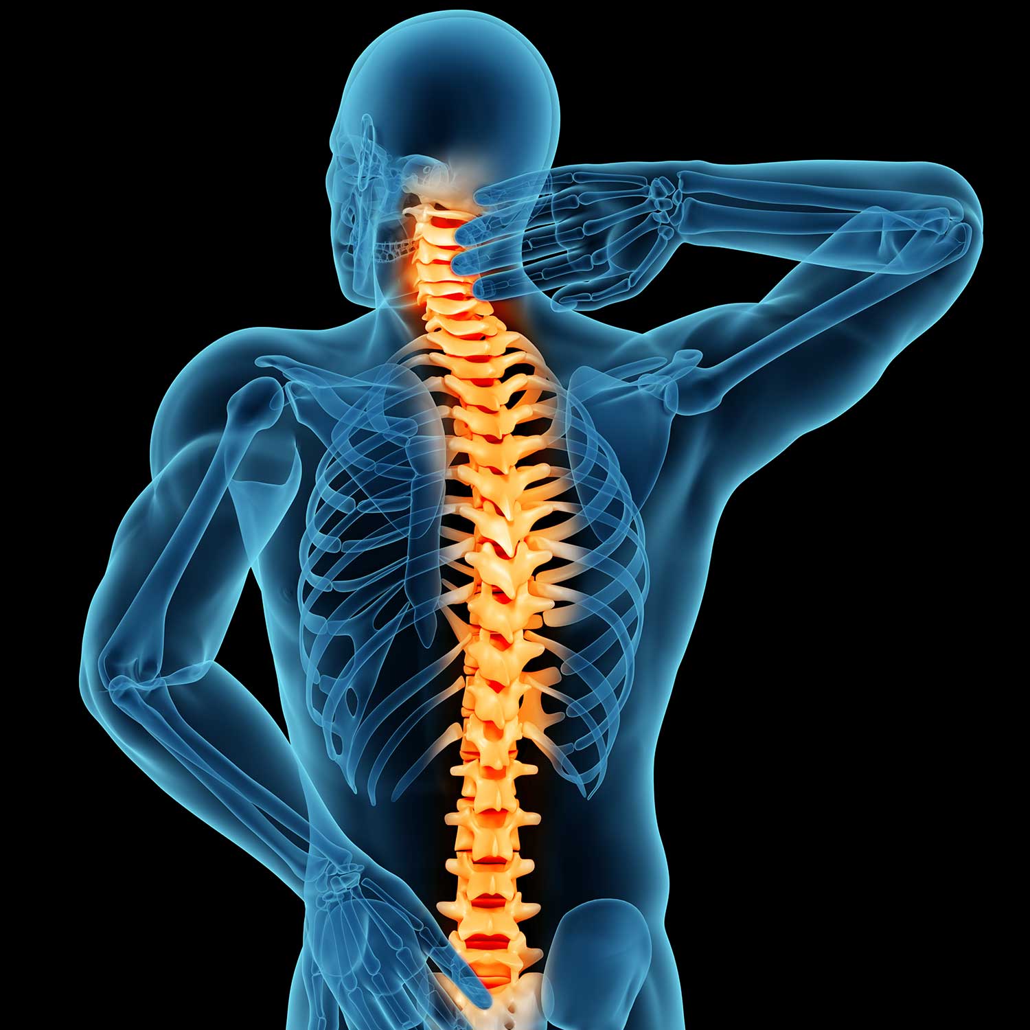
NEWS • Dec 15.2022
Spinal cord pain to PTSD: $5.2m in NHMRC grants to target pressing medical conditions
Both the body and the mind will be a key focus for innovative researchers from the University of Newcastle, who were successful in the latest round of National Health and Medical Research Council (NHMRC) Ideas Grants.
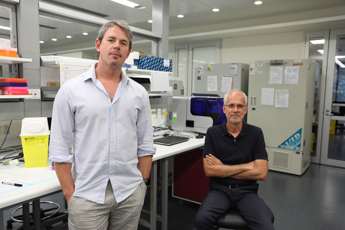
NEWS • Apr 16.2020
Drug repurposing potential for COVID-19
The COVID-19 pandemic has highlighted the need for anti-viral therapies to treat respiratory virus infections. With a vaccine at least 12 months away, drug repurposing offers hope in fast-tracking therapies to treat infected people and save lives.
NEWS • Dec 18.2019
NHMRC awards $9.3 million to 13 University of Newcastle projects
The University of Newcastle has received more than $9.3 million in funding to support projects aiming to solve some of the world’s most critical health problems and improve the lives of millions of Australians.
NEWS • Mar 15.2019
Research underway to improve prostate cancer diagnosis
Funding from the Cancer Council NSW will help advance research in the areas of prostate and lung cancer.
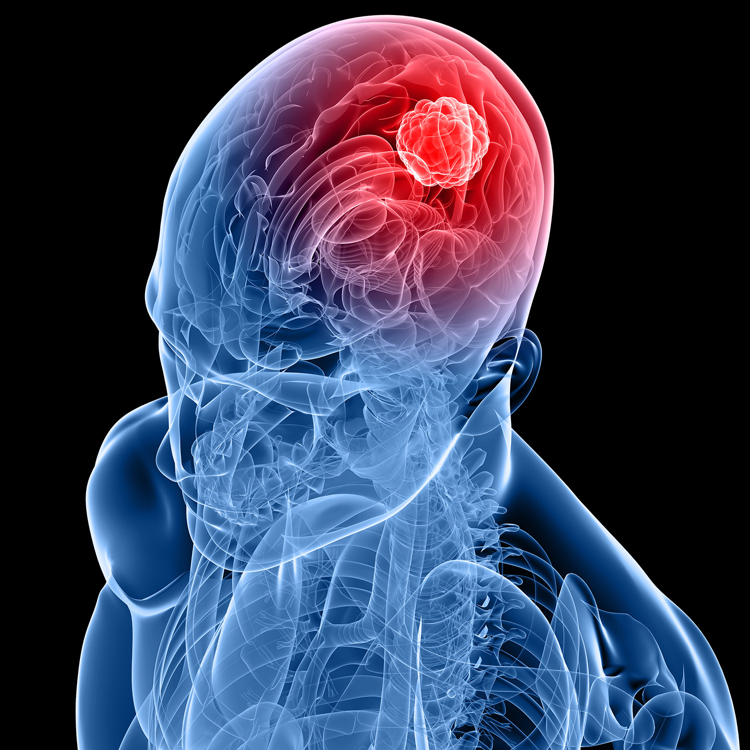
NEWS • Mar 07.2019
Mark Hughes Foundation awards innovation in brain cancer
Five highly innovative brain cancer research projects have received over $725,000 in funding from the Mark Hughes Foundation (MHF) following a nationwide call-out and scientific review by experts in the field.
NEWS • Mar 27.2017
Study strikes a nerve with the spread of cancer
Parallels between tissue regeneration, nerve growth and tumour development, research finds.

NEWS • Oct 23.2013
International spotlight on cancer research
World’s leading cancer researchers will address international Translational Cancer Research Conference in Newcastle.